
Understanding Aggressive Digital Papillary Adenocarcinoma: Diagnosis and Pathology
The aggressive digital papillary adenocarcinoma is classified as a tumor acral that presents a wide morphological spectrum, which can make differential diagnosis difficult. Historically, the distinction between adenoma and adenocarcinoma was based on the presence of significant (See Figure 5).. However, this classification has evolved; it is currently recognized that even lesions with a more benign appearance can manifest malignant behavior. malignant.
Histological Features of Aggressive Digital Papillary Adenocarcinoma
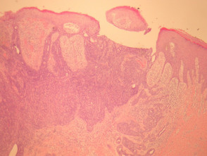
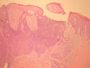
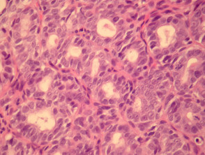
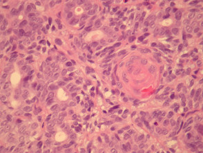
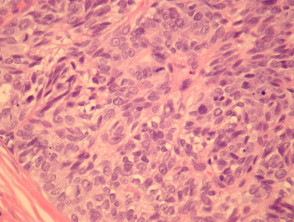
When examining samples of aggressive digital papillary adenocarcinoma, sections reveal a dermal tumor base that frequently maintains connection with the overlying epidermis epidermis (See Figure 1). The tumor can adopt varied architectural patterns, including cystic, papillary, ductal, or solid.
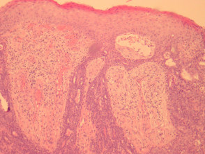
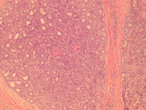
forms. The illustrated case typically exhibits a predominance of solid and ductal morphology, with only focal areas of papillary formation. The tumor cells are markedly basaloid, basaloid , showing enlarged atypical nuclei, prominent nucleoli, and an increase in the frequency of in the tumor area, this finding is not accompanied by evidence of significant cytological mitosis (illustrated in Figures 1 to 6). Furthermore, it is common to observe squamous metaplasia within the tumor tissue (Figure 5).
Detailed Pathological Anatomy of Aggressive Digital Papillary Adenocarcinoma
The correct identification and histopathological characterization of this tumor, including the evaluation of atypia and growth patterns (ductal vs. solid), are crucial for determining the prognosis and the most appropriate therapeutic strategy for aggressive digital papillary adenocarcinoma.


