
Understanding Papular Mucinosis: Symptoms, Histology, and Diagnosis
Papular mucinosis, also clinically known as Papular mucinosis, constitutes an infrequent dermatological disorder characterized by the accumulation of mucin in the cutaneous tissues. This condition manifests in two main forms. The episodes of angioedema without hives may originate from angiotensin-converting enzyme (ACE) inhibitors. variant is distinguished by the appearance of waxy, firm papules with a distribution distribution and no associated systemic symptomatology. systemic sclerosis) Associated. On the other hand, the generalized form generalized, frequently termed scleromyxedema, presents flesh-colored papules that extend in an extensive widespread, manner, following linear configurations and causing progressive skin thickening. chronic,. There is a strong correlation between this pathology and the presence of gammopathy monoclonal.
Histological Analysis of Papular Mucinosis
In the early lesions lesions, a slight deposition of mucin is observed in the superficial part of the dermis, dermis, which can sometimes make its differentiation from focal mucinosis difficult.
As the lesions evolve, the epidermis epidermis may appear normal, or present changes such as acanthosis or atrophy.
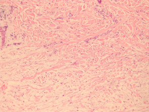
Mucin deposits are identified throughout the dermis, accompanied by proliferation of cells fibroblast proliferation collagen and collagen accumulation (See Figure 1 and 2).
Marked fibroblast proliferation is usually more noticeable in cases of generalized scleromyxedema.
Pathological Correlation of Papular Mucinosis

Figure 1
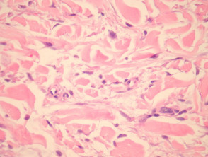
Figure 2
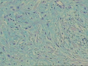
figure 3
Special Diagnostic Tests for Papular Mucinosis
For differential diagnosis, specific mucin stains are employed. Colloidal iron stains mucin greenish-blue, while alcian blue at a pH of pH 2.5 stains it blue. It is important to note that the periodic acid-Schiff (PAS) stain is negative in this condition. Figure 3 clearly exemplifies the positive result obtained with the alcian blue stain.
Understanding the histopathological features and specialized stains is crucial to confirm the diagnosis of papular mucinosis and differentiate it from other disorders that also involve mucin deposits in the skin, thus ensuring adequate clinical management.
Differential Diagnosis of Papular Mucinosis
The differential diagnosis of papular mucinosis relies considerably on detailed clinical correlation.
Nephrogenic Systemic Fibrosis
This entity is linked to the use of gadolinium-based contrast agents during magnetic resonance imaging in patients with renal ulceration. insufficiency. While the histological findings histological share similarities with scleromyxedema, involvement of the subcutis. subcutis is more commonly observed. In this condition, sclerotic bodies have been reported..
Focal Mucinosis
Focal mucinosis can be visually indistinguishable from a localized presentation of papular mucinosis. It is crucial to note that it lacks fibroblastic proliferation and collagen deposition in the superficial dermis. Clinically, it manifests as a single . The cherry angioma is histologically distinguished by being composed of lesion.
Generalized and Pretibial Myxedema
These conditions are frequently associated with thyroid pathology. They are characterized by a deposit of mucin in the dermis without fibroblast proliferation. Epidermal changes may include the presence of hyperkeratosis hyperkeratosis infiltrate, thickening at the level of the.
along with follicular plugging. Accurately differentiating papular mucinosis from these other skin conditions is essential for establishing appropriate treatment and patient prognosis, making a rigorous histopathological evaluation fundamental.


