
Histological Aspects and Diagnosis of Orofacial Granulomatosis
Orofacial granulomatosis, also known as granulomatous cheilitis, clinically manifests as chronic or recurrent swelling of the lips and oral mucosa. This condition requires a detailed analysis of its tissue characteristics.
Histology of Orofacial Granulomatosis
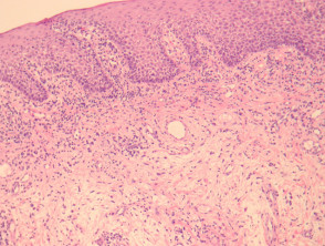
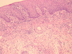
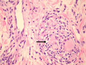
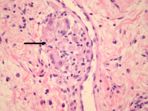
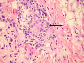
In the histological study of orofacial granulomatosis, oral mucosa samples reveal a sparse inflammatory infiltrate and mild edema (Figure 1). Upon examination at higher magnification, ectasia of the lymphatic vessels is observed alongside isolated and scattered granulomas (Figures 2-4, arrows indicate the location of the granulomas). Occasionally, given the focal nature of the granulomas, their detection can be complex, which is why performing an extensive serial sectioning of the biopsy is frequently recommended to ensure an accurate diagnosis.
Illustrated Pathology of Orofacial Granulomatosis
Special Studies and Microbiology in Orofacial Granulomatosis
Due to the granulomatous appearance of the infiltrate, it is essential to perform special stains for microorganisms. These studies help rule out the presence of underlying infections that could mimic the clinical or pathological presentation of orofacial granulomatosis.
Key Differential Diagnosis of Orofacial Granulomatosis
The differential diagnosis is crucial, as several conditions can present with overlapping histopathological features. It is essential to correlate laboratory findings with the patient's overall clinical presentation.
- Melkersson-Rosenthal Syndrome: It presents with histological pathology identical to orofacial granulomatosis; however, clinically it is distinguished by the concomitant presence of facial nerve paralysis and a fissured (or scrotal) tongue.
- Crohn's Disease: When Crohn's disease affects the oral cavity, the histology may be indistinguishable from orofacial granulomatosis. Therefore, an exhaustive clinical correlation is required to establish the definitive diagnosis.
- Sarcoidosis: Sarcoid granulomas are usually more conspicuous and larger. Nevertheless, there are atypical cases where differentiation from orofacial granulomatosis can be extremely difficult or impossible based solely on histological study.
A thorough evaluation of the histology, along with complementary studies, is essential for an accurate diagnosis of orofacial granulomatosis.


