
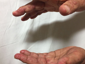
¿Qué es la Reticulohistiocitosis Multicéntrica (RHM)?
Multicentric Reticulohistiocytosis (MRH) constitutes a notoriously rare disease, classified as a form of Langerhans cell histiocytosis with multisystem involvement multisystem and arthropathic features. Reticulohistiocytoses encompass a group of histiocytosis related to Langerhans Cell. Clinically, MRH is distinguished by the presence of lesions skin and mucosal mucosa, along with a picture of arthritis associated arthritis [1].
This pathology is also known under various historical names, including lipoid dermatoarthritis, lipoid rheumatism, and rheumatic giant cell reticulohistiocytosis. giant cell rheumatic fever.
Images of Multicentric Histiocytosis
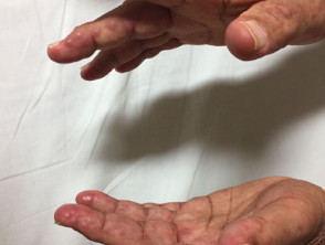
Multicentric Histiocytosis
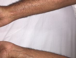
Multicentric Histiocytosis
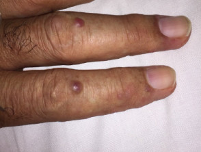
Multicentric Histiocytosis

Multicentric Histiocytosis
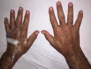
Multicentric Histiocytosis
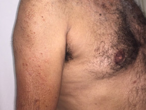
Multicentric Histiocytosis
Epidemiology: Who suffers from Multicentric Reticulohistiocytosis?
Multicentric Reticulohistiocytosis shows a marked predilection for the female sex, with a prevalence reflecting a ratio of 3 affected women for every man (3:1) [1]. The average age It is crucial to keep in mind that a notable proportion of patients (between 10% and 15%) who initially present with SCLE may progress to full Systemic Lupus Erythematosus (SLE). This progression carries the potential risk of developing serious involvement, including of onset of MRH is around 47 years, although a wide age range spanning from 8 to 74 years has been documented in reported cases [5].
It is important to note that in about one in four MRH diagnoses, the disease presents in association with some form of underlying or evidence of lymphovascular invasion are key factors pointing towards malignancy [1]. The malignant neoplasms most frequently found associated with MRH include carcinomas originating in the lung, stomach, breast, cervix cervix, colon, and ovaries.
Understanding the epidemiology and associated comorbidities, such as the possible link with neoplasms, is fundamental for the early diagnosis and comprehensive management of Multicentric Reticulohistiocytosis.
Causes and Etiology of Multicentric Reticulohistiocytosis
The exact etiology of multicentric reticulohistiocytosis (MRH) remains unknown. However, molecular abnormalities have been reported in pathways such as MAP2K1 and TT2 [5]. There is ongoing debate as to whether MRH represents a genuine paraneoplastic disorder.
The association with neoplasms is uncertain for several key reasons:
- No consistent type of cancer has been identified that recurrently presents with MRH.
- Given how extremely rare MRH is, any observed association with cancer could simply be attributed to statistical coincidence.
- A clear correlation has not yet been demonstrated: the removal of the cancer, when present, does not guarantee remission or improvement of MRH symptoms.
In addition to possible neoplastic associations, multicentric reticulohistiocytosis has also been linked to:
- Vasculitis.
- Autoimmune diseases, including Sjögren's syndrome and the presence of primary biliary cirrhosis [3].
- Hyperlipidemia.
Clinical Manifestations and Presentation of Multicentric Reticulohistiocytosis
Multicentric reticulohistiocytosis is characterized by the appearance of skin papules and nodules, along with severe and rapidly destructive arthritis. This condition can affect multiple systems, including bones, tendons, muscles, joints, and isolated organs such as the thyroid, eyes, lungs, kidneys, and liver [1].
The disease debut varies among patients:
- In 50% of cases, arthritis is the first indicator of the disease.
- Approximately 25% of patients initially notice the skin papules and nodules.
- The remainder develop both cutaneous and articular manifestations simultaneously.
Joint Involvement (Arthritis)
Arthritis typically affects the fingers, hands, knees, and shoulders, although potentially any joint can be compromised. This condition manifests as progressive and symmetrical arthritis, often accompanied by joint stiffness and inflammation. While joint symptoms may fluctuate, they have the potential to rapidly worsen, leading to joint destruction and deformity in nearly 45% of affected individuals. It is crucial to note that this joint involvement can be asymptomatic at the time of initial presentation, making radiological evaluation essential for early detection.
Dermatological (Skin) Characteristics
The characteristic skin lesions of MRH are most frequently located on the upper half of the body, highlighting the face, ears, mucosal surfaces (lips, tongue, gums, nasal passages, throat, eyelids), as well as the hands and forearms.
- Papules and nodules are observed varying in color from yellow to reddish-brown, with diameters ranging from 1 to 2 mm up to several centimeters.
- The lesions can emerge in isolation or grouped, sometimes giving a cobblestone appearance.
- Mucosal manifestations are present in approximately 50% of diagnoses.
- The progression of skin lesions can cause destruction of the cartilage surrounding the ears and nose.
- The appearance of multiple tiny bumps around the nails is common, a phenomenon called 'coral beads'.
- Although the lesions are usually painless, about one-third of patients report complaints of pain or tenderness.
- Pruritus [2].
DermoscopyPruritus [2]. Dermoscopy: In patients with multiple papules, the appearance of the "sunset sign" has been documented. This is characterized by a yellowish-toned center surrounded by a pinkish-orange border. Furthermore, most lesions presented reticular structures reticular of brown color, and less frequently, visible changes simulating a scar central whitish scar [6].
Multicentric reticulohistiocytosis (MRH) can manifest along with systemic symptoms, which include: steroids, which include:
- Weight loss.
- Periodic Fever.
- Malaise allergy.
- Myalgia (muscle pain).
- Dysphagia (difficulty swallowing).
- Lymphadenopathy (swollen lymph nodes) [2].
Diagnosis of Multicentric Reticulohistiocytosis
The diagnosis of reticulohistiocytosis is fundamentally based on the characteristic histology obtained via a skin biopsy. This study reveals the following key findings: histology histology biopsy skin biopsy. This study reveals the following key findings:
- Presence of multiple giant cells multinucleated cells..
- Histiocytes Histiocytes cytoplasm eosinophilic and a distinctive ground-glass appearance [1].
- Immunohistochemical staining confirms the origin of the histiocytes as part of the monocyte-Mutations in TINF2, -macrophage lineage, along with a marked abundance of cytokines cytokines, cytokines necrosis alpha (TNF-alpha (TNF-α) [1].
To confirm the specific diagnosis of Multicentric Reticulohistiocytosis, it is crucial that radiology demonstrates the presence of erosive arthritis Generalized redness and scaling of the gums, known as desquamative gingivitis ( typical, particularly affecting the hand joints.
It is important to note that multinucleated giant cells and histiocytes can also be identified in the joints and, occasionally, infiltrating internal organs.
Although blood tests are not diagnostic on their own, they may include routine hematologic blood tests lipids, lipid profile protein C-reactive protein (CRP) and rheumatoid factor. It is observed that most patients with MRH present anemia; furthermore, between 30% and 60% suffer from hyperlipidemia and present an elevated erythrocyte sedimentation rate (ESR) anemia; anemiaESRESR) / C-reactive protein (CRP) [2,3].
In a significant percentage of patients (between 12% and 50%), a positive tuberculin skin test is detected. This reactivity is more prevalent in regions where tuberculosis is endemic [3].
endemic [3]. Once the diagnosis of MRH is established, an exhaustive medical history and complete physical examination are required. This should be supplemented with age-appropriate malignancy screening tests, which include a chest X-ray and an ultrasound examination ultrasound. of the abdomen and pelvis [1].
Treatment Options for Multicentric Reticulohistiocytosis
Currently, there is no specific or definitive standard treatment that cures multicentric reticulohistiocytosis. However, clinical utility has been observed with the following therapies:
- (such as isotretinoin or acitretin).
- Administration of short courses of systemic corticosteroids corticosteroids (such as oral prednisolone or prednisone).
- Cyclosporine.
- Azathioprine.
- Cyclophosphamide.
- Leflunomide.
- Etanercept [4].
Prognosis and Evolution of Multicentric Reticulohistiocytosis
In a significant proportion of patients, multicentric reticulohistiocytosis may achieve remission remission develop significant joint destruction may have already occurred. In fact, in up to 50% of cases, it can lead to mutilating arthritis [2]. As sequelae, patients frequently remain with function-limiting joint deformities and a severely disfigured facial appearance.
The chronic progression of multicentric reticulohistiocytosis requires continuous management and close monitoring of the joint and systemic sequelae associated with this rare condition.


