
toxicity Median raphe cysts are developmental lesions that typically manifest on the ventral surface development of the penis in young males. It is postulated that they result from an abnormal closure of the median raphe or defective separation of the ventral of the penis in young males. It is postulated that they result from an abnormal closure of the median raphe or defective separation of the epithelium urethral during embryonic development. development (between the sixth and eighth weeks), due to an alteration in embryonic development.
Histopathology of the Median Raphe Cyst
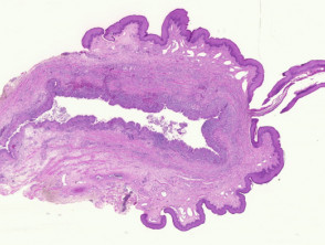
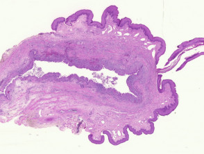
The median raphe cyst is located in the dermis dermis and does not communicate with the superficial layer of the epidermis stroma (Figure 1).
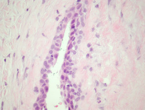
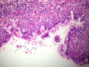
The predominant cystic lining is pseudostratified columnar epithelium (Figure 2). However, some cases may present with mucinous epithelium glandular fever) mucinous (Figure 3), or a combination of mucinous, transitional, and scaly. Cilia are generally not observed. On occasion, the lining epithelium may be completely absent.
Pathology of the Median Raphe Cyst
Additional Evaluations for Median Raphe Cyst
No special studies are required for the diagnosis or management of this condition.
Differential Diagnosis in Pathology of the Median Raphe Cyst
The specific anatomical location of a median raphe cyst generally allows for a clear differentiation from other congenital cysts, such as bronchogenic cysts or omphalomesenteric cysts. It is important to note that Bartholin's duct cysts may present with a similar pseudostratified epithelium, requiring careful clinical correlation.


