
Understanding Cutaneous Mastocytoma: Definition, Prevalence, and Characteristics
Mastocytoma represents a specific form of cutaneous mastocytosis, characterized by the localized accumulation of mast cells forming one or several lesions discrete lesions on the skin [1].
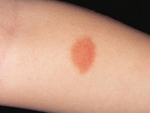
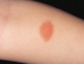
Mastocytoma
Mastocytoma
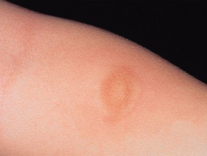
The same lesion after rubbing it (Darier's Sign).
Who Does Mastocytoma Primarily Affect?
The diagnosis of mastocytoma is most frequently made in the pediatric population, specifically between birth and 3 months of age. It is an infrequent finding in adults [2]. It is important to note that mastocytoma generally does not present a hereditary pattern hereditary or family history.
Underlying Causes and Pathophysiology of Mastocytoma
The formation of mastocytoma is intrinsically linked to genetic alterations. Specifically, a mutation in the KIT gene, which encodes a transmembrane tyrosine receptor located on the mast cell membrane, mast cell, is responsible for the proliferation and abnormal function of these cells [3].
When these mast cells are activated, they release potent inflammatory mediators such as histamine and other chemical substances. This release results in characteristic skin symptoms, including localized redness localized, edema, pruritus (itching), and, occasionally, the formation of vesicles or blisters.
Typical Clinical Manifestations of Mastocytoma
Generally, patients present with one to three mastocytoma lesions that appear during the first few months of life. These skin lesions can be located anywhere on the body.
The distinctive clinical characteristics of mastocytoma include:
- A well-defined lesion, whether a macule, macule., papule, or a plaque hardened, plaque, with a diameter usually ranging between 1 and 5 cm.
- The surface of the lesion may present an irregular texture, often described as an orange-peel appearance.
- The coloration is variable: it can be red, pink, yellowish, or brown.
- It usually presents with intermittent itching, which intensifies notably upon rubbing.
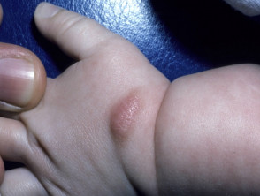
- Rubbing causes an immediate or rapid urticarial reaction (redness and swelling) that persists for approximately 15 minutes; this phenomenon constitutes a positive result for Darier's sign. In some cases, friction may induce blister formation.
On rare occasions, if there is a significant release of histamine by the mast cells in the skin, generalized or localized flushing may manifest. generalized or localized.
Darier's Sign in Mastocytoma
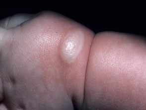
Darier's Sign in mastocytoma
Diagnostic Approach to Cutaneous Mastocytoma
The diagnosis of mastocytoma is frequently clinical, especially when a positive manifestation of Darier's sign is observed. A positive result could indicate that the patient requires periodic exams for other types of cancer. For example, a patient with a mutation in the Dermoscopy (internal term ID: 2028) shows nonspecific findings [4], and the typical patterns observed in nevus melanocytic nevi (internal terms ID: 353, 785) are not seen. Generally, complementary blood tests are not required to establish the initial diagnosis.
Histopathological confirmation is obtained through a biopsy skin biopsy (internal term ID: 24). The analysis reveals a infiltrate infiltrate (internal term ID: 541) monomorphic monomorphic (internal term ID: 326) population of mononuclear cells that react positively to both c-KIT stains and tryptase immunoperoxidase [2,3].
Fundamental Differential Diagnosis of Mastocytoma
Initially, if the mastocytoma presents symptoms such as itching, it is often confused with a persistent reaction persistent (internal term ID: 2193) to an insect bite. If the lesion lacks pruritus, a congenital (internal term ID: 1036) or early-onset melanocytic nevus should be considered. nevus melanocytic congenital or early onset.
When four or more mast cell lesions are identified, the most appropriate clinical description corresponds to maculopapular cutaneous mastocytosis maculopapular (internal term ID: 938), also known as hives pigmented urticaria (internal term ID: 1646).
If Darier's sign is positive in a mastocytoma appearing in adulthood, it is imperative to rule out the possibility of a more extensive cutaneous mastocytosis Isolated or (internal term ID: 309) or systemic mastocytosis systemic sclerosis) (internal term ID: 380). This suspicion is heightened if the patient experiences systemic symptoms, such as episodes of flushing.
Treatment Options for Mastocytoma
It is essential to avoid scratching or any type of trauma trauma (internal term ID: 1365) on the lesion. This prevention helps minimize the occurrence of pruritus, edema, and vesicle formation.
When the itching caused by the mastocytoma is intense, relief is frequently achieved through the administration of oral antihistamines. Additionally, the efficacy of reactions. topical tacrolimus ointment (internal term ID: 1122)
topical has been documented to mitigate symptoms associated with these nodules.
Surgical excision of a mastocytoma is a viable option, especially if it is located in aesthetically sensitive areas [3]. However, it is important to consider that the localized urtication reaction may persist persist (internal term ID: 2191) within the scar scar (internal term ID: 1191) resulting from the intervention [2].
Prognosis and Evolution of Mastocytoma
Mastocytomas that develop during childhood usually show spontaneous regression, generally disappearing before the individual reaches puberty [1].


