
Lymphangioma Circumscriptum: Clinical and Histopathological Analysis
Lymphangioma circumscriptum manifests visibly on the skin surface as clusters of thin, translucent, lymph-filled vesicles, resembling bunches of grapes or frog spawn. The presence of bleeding within these lesions can alter their coloration to dark red or black tones.
Detailed Histopathology of Lymphangioma Circumscriptum
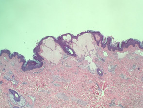
Histopathological examination of lymphangioma circumscriptum reveals the presence of acanthosis and hyperkeratosis in the epidermis (Figure 1). In the papillary and reticular dermis, dilated lymphatic channels containing eosinophilic proteinaceous material are observed in the papillary dermis (Figures 2 and 3).
Pathological Images of Lymphangioma Circumscriptum
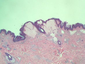
Figure 1
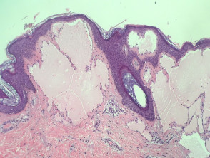
Figure 2
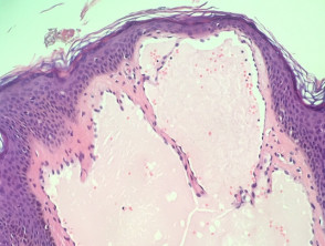
Figure 4
Generally, no additional histochemical staining is required. However, lymphatic architecture can be effectively highlighted using specific immunohistochemical markers such as D2-40.
Differential Diagnosis of Lymphangioma Circumscriptum
Angiokeratoma can present a clinical appearance very similar to lymphangioma circumscriptum. The key distinction lies in the vascular composition: while lymphangioma contains lymph-filled lymphatic vessels, angiokeratoma is composed of blood vessels harboring blood.


