Melanoma in Situ: Visual Guide and Examples
Melanoma In Situ
Melanoma In Situ
Melanoma In Situ
Melanoma In Situ
Melanoma In Situ
Melanoma In Situ
Melanoma In Situ
Melanoma In Situ
Melanoma In Situ
Melanoma In Situ
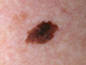
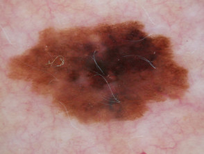
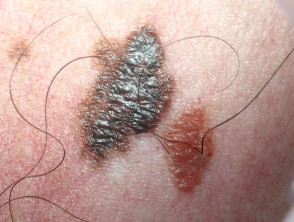
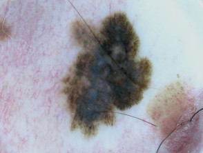
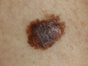
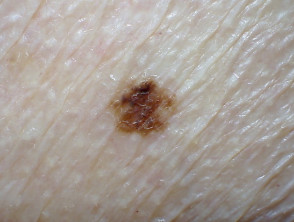
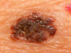
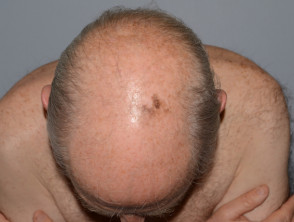
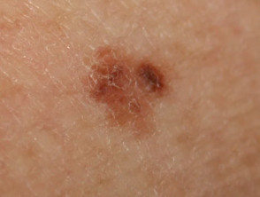
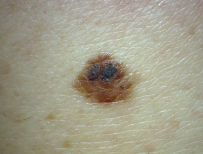
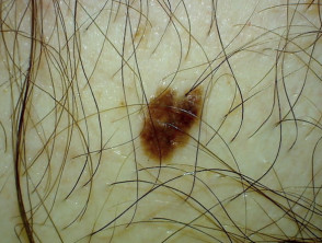
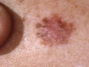
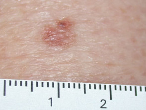
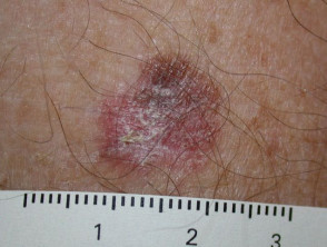
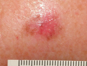
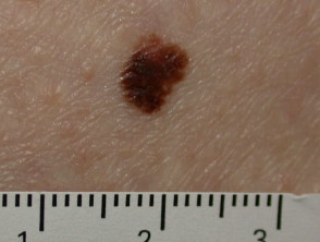
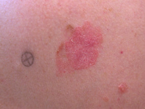
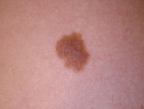
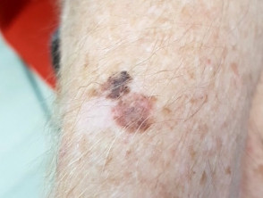
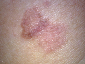
The image collection presented here offers an essential visual resource for understanding and distinguishing Melanoma in Situ, the earliest stage of skin cancer originating in keratinocytes. Recognizing these visual characteristics is fundamental for early intervention and treatment success.
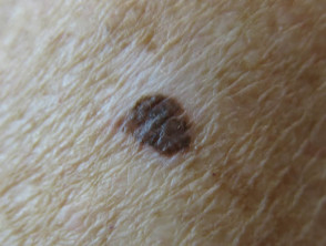
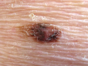
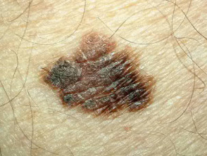
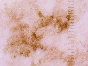
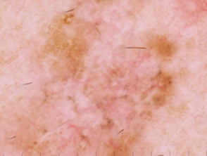
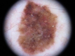
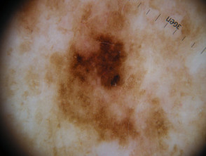
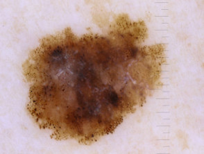
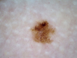
In Situ Dermatoscopy of Melanoma
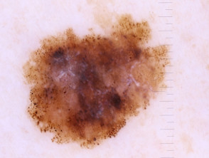
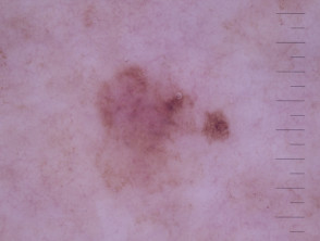
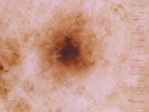
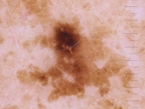
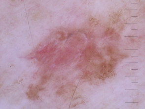
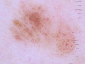
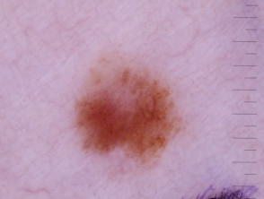
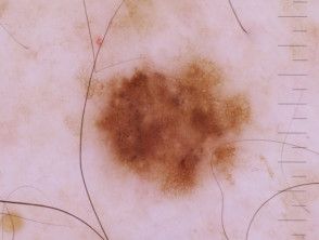
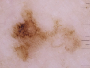
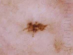
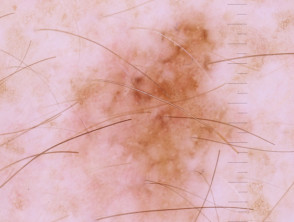
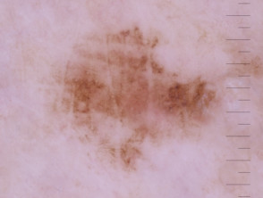
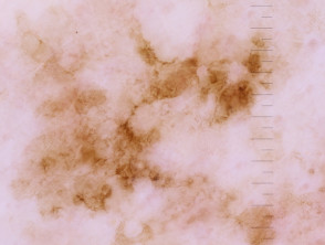
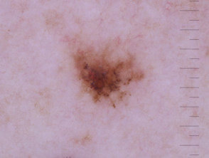
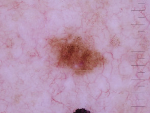
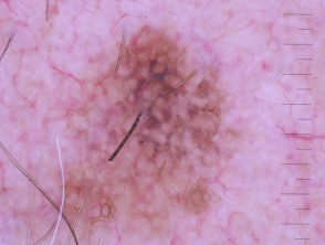
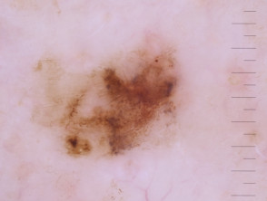
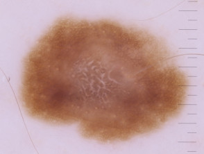
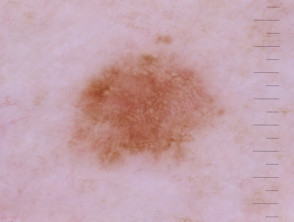
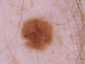
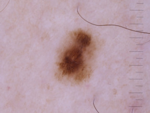
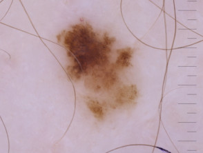
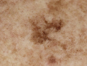
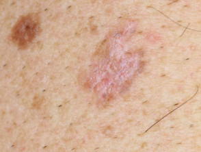
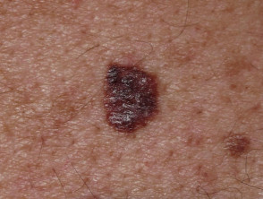
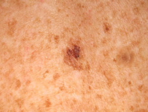
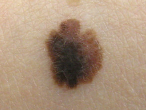
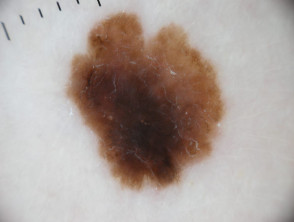
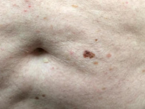
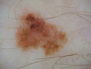
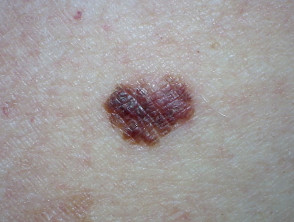
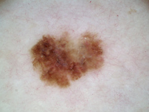
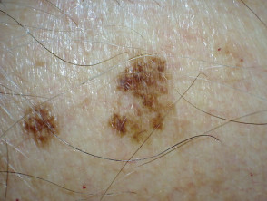
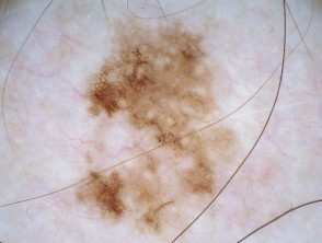
Comparative Images: Macroscopy and Dermatoscopy of Melanoma In Situ
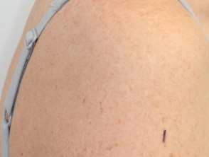
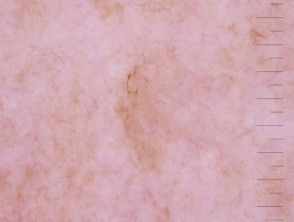
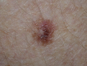
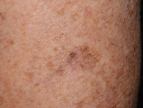
The analysis of melanoma in situ (MIS) cases requires precise correlation between the macroscopic image and dermatoscopic visualization. This comparison is fundamental for identifying atypical patterns characteristic of melanocytic neoplasia in its initial stages.
Next, we will explore the importance of these image pairs for differential diagnosis and confirmation of a melanoma that has not yet invaded the dermis. Dermatoscopy offers a diagnostic depth unattainable to the naked eye, revealing key structural and pigmentary characteristics.
Visual Correlation in Dermatoscopic Diagnosis of Melanoma
The correct identification of dermatoscopic components, such as atypical pigmentary network, the presence of globular atypia, or irregular vascular patterns, allows distinguishing melanoma in situ from benign lesions like solar lentigines or atypical nevi. The image collection presented above illustrates the morphological variability of MIS.
For a comprehensive dermatoscopic diagnosis, a structured mnemonic rule, such as the ABCDE system or more advanced criteria, must always be applied to systematically evaluate every suspicious lesion. Mastering this macro-microscopic correlation is an essential pillar in the early detection of skin cancer.