Images of Melanocytic Nevi in Skin of Color

type1__protectwyjqcm90zwn0il0_focusfillwzi5ncwymjisinkildy2xq-5000872-6735090-jpg-9379786

Table of Contents

Go to the image library

Fitzpatrick Skin Phototype I: Characteristics and Visualization

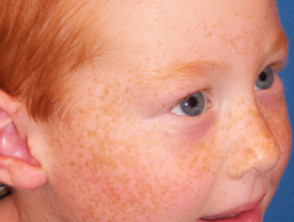
Phototype I on the Fitzpatrick scale represents the lightest and most sun-sensitive skin. People with this skin type usually have a very fair complexion, often with freckles, blonde or red hair, and light eyes. This skin has one fundamental characteristic: it never tans; it only burns easily.
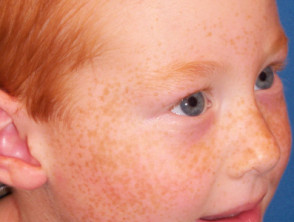
Visual representation of skin phototype 1.
Skin Phototype 1
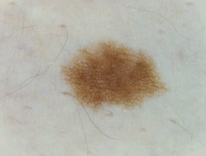
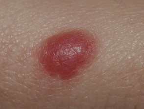
Classic Spitz nevus observed on phototype 1 skin.
Spitz nevus, skin type 1
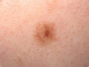
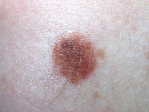
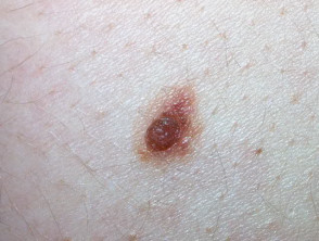
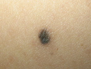
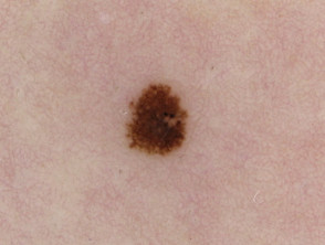
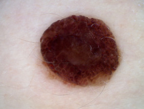
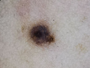
Macroscopic image of a common nevus on type 1 skin.
Nevus, skin type 1, macro
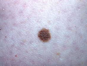
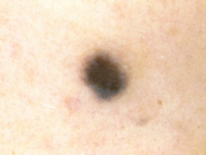
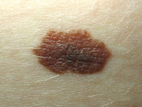
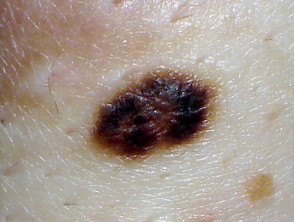
Detail of a nevus on type 1 skin.
Nevus, skin type 1, macro
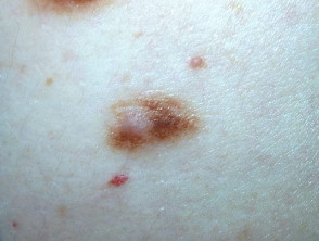
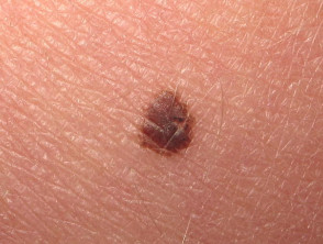
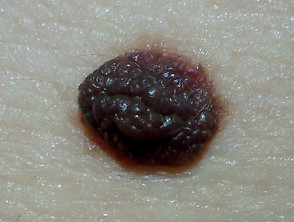
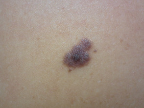
Another detailed view of a common nevus on Fitzpatrick I skin.
Nevus, skin type 1, macro
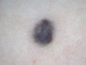
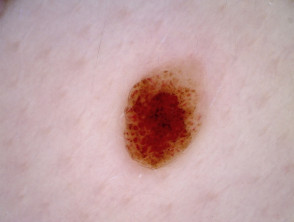
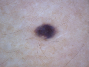
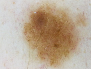
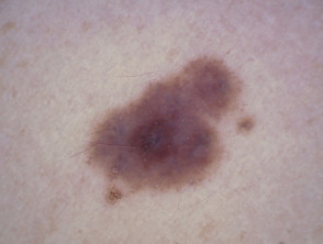
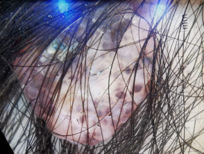
Dermatoscopy of a blue nevus in a patient with type 1 skin.
Blue nevus, skin type 1 dermatoscopy
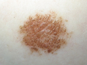
Dermatoscopy of a Spitz pigmented nevus on Fitzpatrick I skin.
Spitz pigmented nevus, skin type 1 dermatoscopy
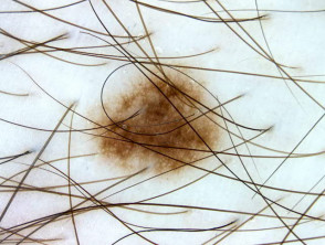
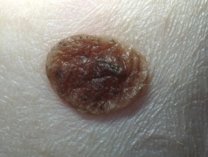
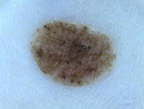
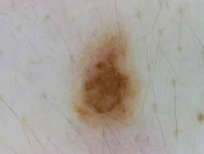
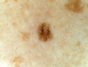
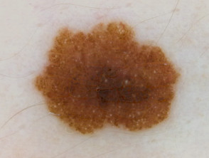
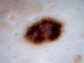
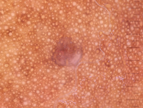
Dermoscopic visualization of a nevus on type 1 skin.
Nevus, skin type 1, dermatoscopy

Dermoscopic Analysis: Fitzpatrick Skin Type 1 (Macro and Dermatoscopy)

The evaluation of pigmented lesions in phototype I requires special attention due to the high incidence of sunburns and poor basal pigmentation. Below are comparisons between the macroscopic image and the dermoscopic analysis of specific nevi in this skin type.
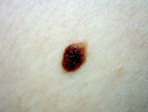
Macroscopic image of a nevus on type 1 skin, reference 1

Nevus skin type 1, macro 1

Dermoscopic analysis of the nevus, skin type 1, reference 1

Nevus, skin type 1, dermatoscopy 1

Macroscopic image of a second nevus on type 1 skin, reference 2

Nevus skin type 1, macro 2

Dermoscopic analysis of the second nevus, skin type 1, reference 2

Nevus, skin type 1, dermatoscopy 2

Macroscopic image of an immature Spitz nevus on type 1 skin, reference 3

Immature Spitz nevus, skin type 1, macro 3

Dermoscopic analysis of the immature Spitz nevus, skin type 1, reference 3

Immature Spitz nevus, skin type 1, dermatoscopy 3

Macroscopic image of a junctional melanocytic nevus on type 1 skin, reference 4

Junctional nevus, skin type 1, macro 4

Dermoscopic analysis of the junctional melanocytic nevus, skin type 1, reference 4

Junctional nevus, skin type 1, dermatoscopy 4

The management of Fitzpatrick Skin Type I is essentially preventive. Given its inability to generate protective melanin, these patients must rigorously adhere to sun protection measures (clothing, hats, and broad-spectrum sunscreens with high SPF) to mitigate the risk of actinic damage and skin cancer.
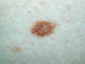
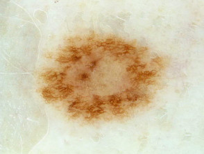
Macroscopic image of a nevus on type 1 skin.
Nevus, skin type 1, macro 2
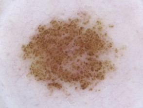
Dermoscopic image of a nevus on type 1 skin.
Nevus, skin type 1, dermatoscopy 2
Macroscopic image of a cobblestone nevus on type 1 skin.
Cobblestone nevus, skin type 1, macro 3
Dermoscopic image of a cobblestone nevus on type 1 skin.
Cobblestone nevus, skin type 1, dermatoscopy 3

Fitzpatrick Skin Types: Analysis of Type 2

Next, we examine the clinical and dermoscopic characteristics associated with Fitzpatrick Phototype 2.
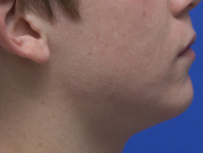
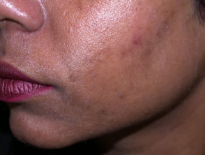
Visual representation of skin phototype 2.
Skin Phototype 2
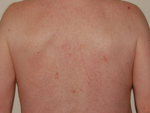
Another visual representation of skin phototype 2.
Skin Phototype 2
Dermatoscopy of a blue nevus diagnosed on type 2 skin.
Blue nevus, skin type 2, dermatoscopy

Macroscopic and Dermoscopic Image Pairs for Fitzpatrick Skin Type 2

Direct comparison between the clinical view (macro) and observation with a dermatoscope is essential for the accurate evaluation of lesions in light phototypes like Type 2.
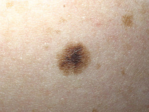
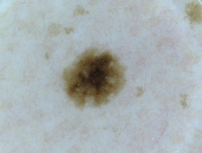
Nevus, skin type 2 in macroscopic view, case 1.
Nevus, skin type 2, macro 1
Nevus corresponding to case 1 seen with a dermatoscope on type 2 skin.
Nevus, skin type 2, dermatoscopy 1
Second case of nevus on type 2 skin, macro view.
Nevus, skin type 2, macro 2
Nevus on type 2 skin observed via dermatoscopy.
Nevus, skin type 2, dermatoscopy 2
Nevus on type 2 skin, macroscopic view.
Nevus, skin type 2, macro 3
Nevus on type 2 skin examined with a dermatoscope.
Nevus, skin type 2, dermatoscopy 3
Nevus on type 2 skin, macro image.
Nevus, skin type 2, macro 4
Nevus on type 2 skin under dermoscopic examination.
Nevus, skin type 2, dermatoscopy 4

Fitzpatrick Skin Types 3

The third phototype on the Fitzpatrick scale is characterized by slightly darker pigmentation, which significantly influences the skin's response to solar radiation and dermatological procedures.
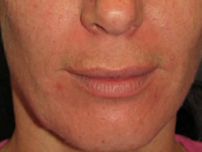
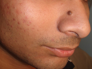
Representation of a person with skin phototype 3.
Skin Phototype 3
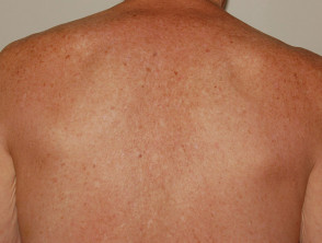
Visual example of skin phototype 3.
Skin Phototype 3
Melanocytic nevus on type 3 skin, macro view.
Nevus, skin type 3, macro
Melanocytic nevus on type 3 skin examined with a dermatoscope.
Nevus, skin type 3, dermatoscopy

Comparative Analysis: Dermoscopic and Macroscopic Images of Fitzpatrick Skin Type 3

Below are specific examples illustrating the characteristics of nevi and the appearance of skin classified as Fitzpatrick type 3 under different magnifications.
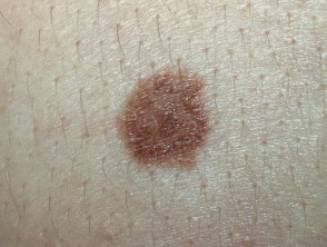
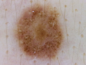
Nevus on type 3 skin, detailed macroscopic image.
Nevus, skin type 3, macro 1
Nevus, skin type 3, dermatoscopy 1
Blue nevus, skin type 3, macro 2
Blue nevus, skin type 3, dermatoscopy 2
Nevus, skin type 3, macro 3
Nevus, skin type 3, dermatoscopy 3
Nevus, skin type 3, macro 4
Nevus, skin type 3, dermatoscopy 4

Fitzpatrick Phototypes 4

The following section explores the specific characteristics associated with phototype IV on the Fitzpatrick scale, which is fundamental for understanding skin responses to sun exposure.
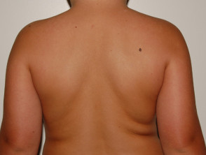
Skin Phototype 4
Skin Phototype 4

Comparative Images: Dermatoscopy and Lesions on Fitzpatrick Skin Type 4

Below are image pairs contrasting the macroscopic appearance with dermoscopic findings of nevi in individuals with type 4 skin, facilitating clinical analysis.
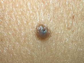
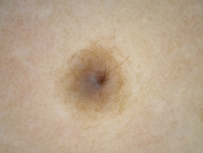
Nevus, skin type 4, macro 1
Nevus, skin type 4, dermatoscopy 1
Nevus, skin type 4, macro 2
Nevus, skin type 4, macroscopic view
Nevus, skin type 4, dermoscopic view
Nevus, skin type 4, additional macroscopic view
Nevus, skin type 4, secondary dermoscopic view
Nevus, skin type 4, third macro view
Nevus, skin type 4 dermatoscopy, final detail

Nevus Characteristics on Fitzpatrick Skin Type 5

Individuals classified with Fitzpatrick phototype V have skin that rarely burns and tans easily, characterized by pigmentation that is already substantially darker than previous types. Examining the behavior and morphology of a nevus on this skin type provides crucial information for differential diagnosis.
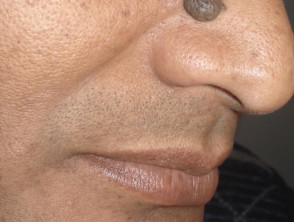
Typical melanocytic nevus on skin phototype 5
Nevus, skin type 5
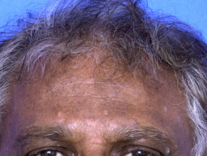
Visual representation of skin phototype 5
Skin Phototype 5
Reference image of skin phototype 5
Skin Phototype 5
Distinctive melanocytic nevus on the face of a person with type 5 skin
Melanocytic nevus, skin type 5
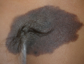
Congenital nevus on type 5 skin with magnified view
Congenital nevus, skin type 5, macro

In phototype V, diagnostic difficulty can vary, as although the pigmentation base is high, pigmented lesions still require meticulous evaluation. It is essential to document any asymmetry, irregular border, or change in color, regardless of the patient's basal skin tone. Accurate documentation of these nevi is key to monitoring their evolution over time and differentiating between benign lesions and those that might require intervention.
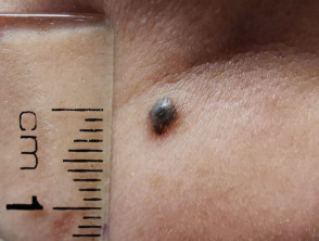
Dermal nevus on type 5 skin, macroscopic view

Dermal nevus, skin type 5, macro

Macroscopic and dermoscopic image pairs of Fitzpatrick skin type 5
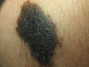
Congenital nevus, skin type 5, macroscopic image 1

Congenital nevus, skin type 5, macro 1
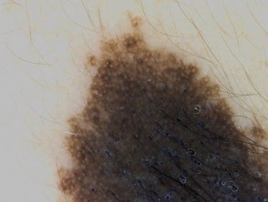
Congenital cutaneous nevus type 5, dermoscopic study 1

Congenital cutaneous nevus type 5, dermatoscopy 1
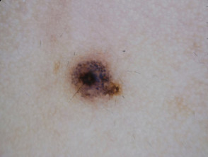
Dermal nevus, skin type 5, macroscopic image 2

Dermal nevus, skin type 5, macro 2

Dermal nevus, skin type 5, dermoscopic study 2

Dermal nevus, skin type 5, dermatoscopy 2
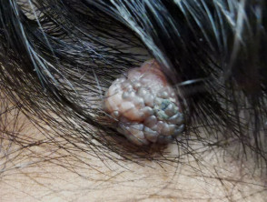
Dermal nevus, skin type 5, macroscopic image 3

Dermal nevus, skin type 5, macro 3

Dermal nevus, skin type 5, dermoscopic study 3

Dermal nevus, skin type 5, dermatoscopy 3

Fitzpatrick Skin Type 6
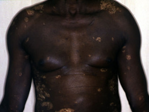
Skin phototype 6, presenting early vitiligo

Skin Phototype 6, showing early vitiligo
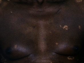
Representation of Fitzpatrick skin type 6

Fitzpatrick Skin Type 6

Nevus on type 6 skin, dermoscopic visualization

Nevus, skin type 6, dermoscopy

Fitzpatrick classification is essential for assessing skin susceptibility to sun damage and the risk of developing pigmented lesions. The detailed visualization of nevi on both type 5 skin—with its dermal and congenital nuances—and type 6 skin underscores the importance of accurate differential diagnosis, supported by techniques like dermatoscopy, regardless of the cutaneous phototype.


Dermatly.com - El sitio de tu piel