
Flegel Disease: Clinical Features and Histopathology
Flegel disease, also known as hyperkeratosis lenticularis perstans, is a dermatosis rare dermatosis characterized by the appearance of asymptomatic papules that is repaired with a transverse suture).. These lesions are predominantly located on the dorsa of the feet and the lower portion of the legs. While the diagnosis is often suggested by clinical findings, confirmation requires rigorous histopathological level. analysis. It is theorized that this disorder originates from alterations in the lamellar bodies (Odland bodies) within the keratinocytes.
Histology of Hyperkeratosis Lenticularis Perstans (Flegel Disease)
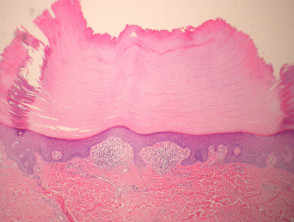
In histological sections corresponding to Flegel disease, well-defined areas of compact lamellar hyperkeratosis are observed, accompanied by focal parakeratosis (see Figure 1). Beneath the hyperkeratotic layer, the epidermis exhibits an atrophic appearance when compared to the surrounding skin. Furthermore, the granular layer granulomatous is thinned or absent (Figure 2). In the dermis papillary dermis, it is common to find a dense infiltrate infiltrate composed mainly of small lymphocytes lymphocytes scattered among and a smaller proportion of histiocytes.. histiocytes. The presence of dilation of superficial vessels is also evident (Figure 3).
Pathological Anatomy of Hyperkeratosis Lenticularis Perstans (Flegel Disease)
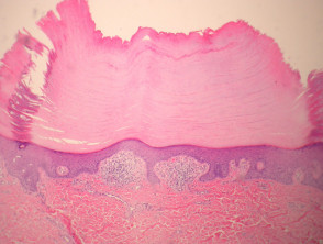
Figure 1
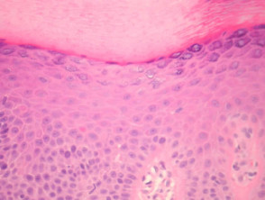
Figure 2
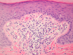
Figure 4
Special Studies for Hyperkeratosis Lenticularis Perstans (Flegel Disease)
Generally, performing additional complementary studies is not required to establish the diagnosis of Flegel disease. Although the examination of lamellar granules via microscopy electron microscopy is frequently mentioned in the literature, this procedure is not considered essential or decisive for confirming the patient's clinical diagnosis.
Confirming the clinical diagnosis via histopathology is fundamental to differentiate Flegel disease from other skin conditions presenting with keratosis. Although the histological findings are characteristic, the prognosis is usually benign, requiring symptomatic management in most cases.
Differential Diagnosis of Hyperkeratosis Lenticularis Perstans (Flegel Disease) in Pathology
Differentiating Flegel Disease from other dermatoses with hyperkeratosis is crucial for an accurate diagnosis. Below are the key conditions to consider in the differential diagnosis:
Kyrle's Disease
Kyrle Disease presents notable clinical similarities to Flegel Disease and may share some pathogenetic elements. However, Kyrle Disease typically manifests a distinctive nail edge keratotic border and Congenital fibromatosis perforating material prominent in the epidermis, findings that help distinguish it.
Porokeratosis
The diagnosis of porokeratosis is significantly facilitated by the presence of strata corneum, a characteristic pathological sign of this condition.
Actinic Keratosis
In actinic keratosis, actinic dysplasia is observed. dysplasia actinic. Some experts suggest that Flegel Disease might represent an early or incipient manifestation of epidermal dysplasia..
Correctly differentiating these entities requires a detailed histopathological evaluation, as the clinical appearance can be misleading, making the analysis of the keratinization pattern and the underlying inflammatory response essential.


