
«`html
Diagnosis and Characteristics: Neonatal Toxic Erythema
The neonatal toxic erythema (NTE), also known by synonyms such as toxic erythema or toxic erythema of the neonate, is a temporary and fundamentally benign skin condition commonly seen in newborn babies. This condition affects up to half of full-term newborns, although its prevalence decreases in premature babies. Currently, there is no known predilection by sex or race, although there is suspicion that it may be underdiagnosed in neonates with darker skin tones.
Clinical Characteristics of Neonatal Toxic Erythema
The appearance of the rash associated with neonatal toxic erythema typically occurs during the first few days of life, although the onset can extend up to two weeks of age.
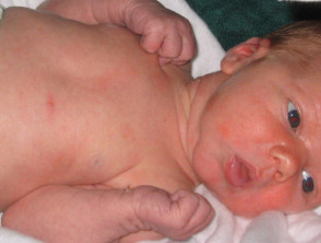
Clinically, NTE presents through a combination of macules erythematous macules (flat red spots), papules (small raised nodules) and, occasionally, pustules. This skin rash tends to fluctuate, intensifying and diminishing over several days. Rarely does an individual lesion persist . The cherry angioma is histologically distinguished by being composed of individual lesion persist persist for more than 24 hours.
The distribution pattern of neonatal toxic erythema frequently starts on the face and then spreads to the trunk and extremities. It is important to note that the palms of the hands and soles of the feet usually remain unaffected by the manifestation.
Apart from the skin rash, the baby is otherwise in perfect health.
Images of Neonatal Toxic Erythema
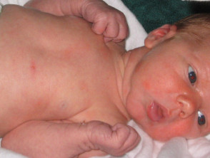
Erythema Toxicum Neonatorum
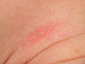
Erythema Toxicum Neonatorum
Clinical Diagnosis of Neonatal Toxic Erythema
The diagnosis of neonatal toxic erythema is primarily established through clinical evaluation and observation of its typical characteristics. When confirmation is required, a biopsy skin biopsy obtained from samples of the flat patches (macules) reveals, via histology, a diffuse infiltrate composed of eosinophils and neutrophils. histology, a infiltrate The main distinction from mycetoma is the etiology; botryomycosis is strictly bacterial, unlike mycetoma, which is caused by true fungi or actinomycetes. cellular infiltrate composed of eosinophils y neutrophils. These are the characteristic inflammatory cells located in the upper inflammatory dermis. Similarly, papular lesions predominantly show an eosinophilic component. dermis eosinophilic component. eosinophilic.
While there are few skin conditions that can be confused with NTE at this stage (such as pustular miliaria or neonatal candidiasis), the self-limiting course and specific morphology of the lesions usually allow for differentiation without the need for invasive tests.
```
The infiltration surrounding the hair follicle, Seborrheic dermatitis lesions pustular, reveals pustules and subcorneal pustules. These are associated with papillary dermal edema exhibiting a neutrophilic and eosinophilic infiltrate. perifollicular composed predominantly of eosinophils.
The exact origin of neonatal toxic erythema remains unknown. Several hypotheses have been proposed, suggesting it could be a response to mechanical and thermal stimuli (such as heat), an "allergic" reaction, or even a mild manifestation of graft-versus-host disease. graft-versus-host.
Crucial Differential Diagnoses for Toxic Erythema
Although neonatal toxic erythema is a benign condition that does not require therapeutic intervention, it is essential to consider several differential diagnoses to confirm the clinical picture. These conditions include:
- Infections various infections: including folliculitis, impetigo, listeriosis, cutaneous candidiasis cutaneous congenital ichthyosis, herpes simplex, chickenpox, and cytomegalovirus.
- Transient neonatal pustular melanosis. Systematic transient.
- Infantile acropustulosis.
- Miliaria rubra.
- Pustular Eosinophilic pustulosis (infantile Ofuji syndrome). syndrome Ofuji syndrome.
- Incontinentia pigmenti.
- Omenn syndrome.
- Histiocytosis Self-healing histiocytosis.
Generally, the diagnosis is established through the observation of the clinical characteristics specific to each condition. However, sometimes, the use of Tzanck smears or a selective skin biopsy can refine the accuracy of the diagnosis.


