
«`html
¿Qué es un Nevus Epidérmico? Definición y Clasificación
An epidermal nevus is characterized by excessive proliferation of the epidermis. These lesions may be present at birth (in 50% of cases) or develop during childhood, mainly in the first year of life. The abnormality arises from a defect in the ectoderm, which is the outer embryonic layer that gives rise to both the epidermis and the neural neural.
Main Types of Epidermal Nevi
Skin lesions commonly classified as epidermal nevi result from the uncontrolled growth of keratinocytes, which are the predominant horny cells in the skin.
- Epidermal Nevus linear
- Epidermal Nevus epidermolytic
- Epidermal Nevus acantholytic (with similarities to Hailey-Hailey disease or Darier's disease)
- Porokeratosis linear
- Segmental epidermal nevi
If another skin component is predominant in the involvement, the . The cherry angioma is histologically distinguished by being composed of is called an organoid nevus.
Genetic Causes and Distribution of Epidermal Nevus
Normally, there are two copies of each gene, one inherited from the mother and one from the father. It is postulated that there are two populations of skin cells, one carrying the maternal gene and another with the paternal gene (a phenomenon known as mosaicism). If one of these skin cell populations presents a mutation, it can manifest as a localized area of thickened skin, constituting an epidermal nevus. It is uncommon for epidermal nevi to affect several members of the same family. Mutations have been identified gene maternal gene and another with the paternal gene (a phenomenon known as mosaicism). If one of these skin cell populations presents a mutation, it can manifest as a localized area of thickened skin, constituting an epidermal nevus. It is uncommon for epidermal nevi to affect several members of the same family. Mutations have been identified mutations in the FGFR3, PIK3CA, and HRAS genes.
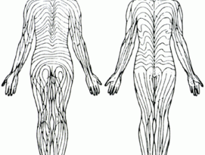
The distribution of epidermal nevi follows Blaschko's lines. These lines represent the trajectories followed by groups of genetically identical cells during embryonic development development. Skin cells carrying the abnormally active gene expand to form the epidermal nevus, while healthy cells form the rest of the normal-appearing skin.
Recent research has identified point mutations in nail edge (keratin) genes, which reinforces this theory. The abnormal gene resides specifically in the cells of the epidermal nevus, but not in the surrounding healthy skin. The same abnormalities in keratin 1 and keratin ten have been detected in both parents affected by epidermolytic epidermal nevus and in their children suffering from Ichthyosis erythrodermic bullous (an infrequent variant of Ichthyosis). Therefore, epidermolytic epidermal nevus is considered a mosaic manifestation of this form of ichthyosis.
Likewise, the mutation of the ATP2A2 gene, associated with Darier's disease, has been found in the affected cells of a patient with acantholytic epidermal nevus. This suggests that this subtype of epidermal nevus could be a mosaic presentation of Darier's disease. Finally, porokeratosis linear could represent a mosaic form of Ichthyosis superficial disseminated ichthyosis.
Understanding the genetic basis and distribution of these lesions helps dermatologists to more accurately classify the specific type of epidermal nevus present and to explore targeted treatments for these complex skin malformations.
```
porokeratosis actinic.
Blaschko's Lines
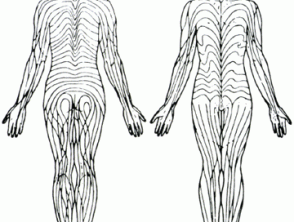
Blaschko's Lines
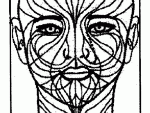
Blaschko's Lines
Characteristics of Epidermal Nevus
Linear Epidermal Nevus
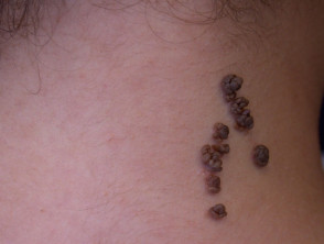
An epidermal nevus typically manifests on the trunk and extremities, being less frequent on the face or scalp. Most cases correspond to linear epidermal nevi, which present as a demarcated band, generally restricted to one side of the body (known as unilateral nevus or unilateral o nevus unius lateralis). At birth or in early childhood, these marks appear flat and tan or brown in color. With advancing age, the lesion tends to thicken and develop a warty appearance warty. Furthermore, the size of the nevus may progressively increase over several years.
Segmental Epidermal Nevi
Segmental epidermal nevi are a less common presentation, sometimes referred to as ichthyosis hystrix. In these cases, multiple lesions are observed that frequently adopt a swirling pattern, extending to one or both sides of the body. In a minority of patients, this type of nevus is associated with other malformations congenital, particularly affecting the skeletal structure and the central nervous system (CNS).
Histopathology of Epidermal Nevus
It is important to note that macular amyloidosis can coexist with lichenoid amyloidosis in up to 25% of diagnosed patients [9]. biopsy Upon performing a skin biopsy, the epidermal nevus reveals a marked thickening of the epidermis. It is useful to review the specific pathology of the epidermal nevus for its classification: pathology specific to the epidermal nevus for its classification:
- The epidermolytic subtype, which is infrequent, is distinguished by a histological pattern histological called hyperkeratosis epidermolytic hyperkeratosis, similar to that seen in bullous congenital ichthyosiform erythroderma.
- The acantholytic epidermal nevus presents pathological findings reminiscent of Darier's disease.
- The linear variant of porokeratosis exhibits pathology resembling superficial disseminated actinic porokeratosis.
Epidermal Nevi
epidermal nevus
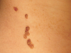
epidermal nevus
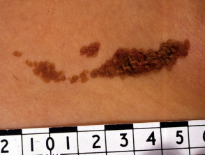
epidermal nevus
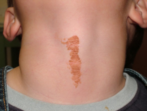
epidermal nevus
Understanding the histology and the linear or segmental nature of epidermal nevi is crucial for the accurate diagnosis of these congenital skin malformations.
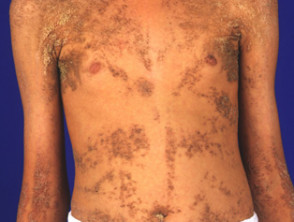
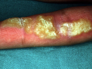
Syndromes Associated with Epidermal Nevi
Epidermal nevus syndromes involve the coexistence of a keratinocytic or organoid epidermal nevus along with abnormalities in other organ systems derived from the embryonic ectoderm. These conditions can manifest by notably affecting the eyes, skeleton, or nervous system. Clinically, numerous specific syndromes have been identified based on these associations.
The underlying alteration that produces the skin lesions can also cause disorders in vital internal organs such as the brain, eyes, and skeleton. When conditions involving multiple organ systems exist, the complex is called an epidermal nevus syndrome (ENS).
Complications of Epidermal Nevi
In most cases, epidermal nevi are stable lesions that persist into adulthood without causing significant complications.
Differential Diagnosis of Epidermal Nevus
Inflammatory linear verrucous epidermal nevus (ILVEN) is frequently considered a separate entity from typical epidermal nevus.
Organoid nevi are characterized by the benign overgrowth benign of epidermal appendage structures, including:
- Congenital Melanocytic Nevus sebaceous
- Sebaceous nevus salivary sweat gland
- Congenital Melanocytic Nevus comedo
- Becker's nevus.
On the other hand, melanocytic nevi melanocytic nevi are defined by the proliferation benign proliferation of nevus melanocytes.
Treatment for Epidermal Nevi
Currently, there is no medical treatment with universally proven efficacy to resolve epidermal nevi. In certain situations, topical calcipotriene topical can help reduce the thickness of the skin lesion. If removal is required, procedures such as surgical excision or laser treatment can be considered on the affected nevi. laser on the affected nevi.
The management of syndromes associated with epidermal nevi requires a multidisciplinary approach, prioritizing the treatment of the involved systemic manifestations along with the dermatological management of the primary skin lesion.


