
Discoid Lupus Erythematosus (DLE): Understanding Causes, Symptoms, and Clinical Prevalence
Lupus erythematosus (LE) encompasses a complex spectrum of diseases persistent. It commonly affects smokers and is considered pre, inflammatory y are evaluated in patients with suspected connective tissue or autoimmune disease., with the skin being one of the frequently affected areas. In particular, discoid lupus erythematosus (DLE) is established as the predominant chronic manifestation of cutaneous lupus cutaneous. It is clinically distinguished by the emergence of plaques cells scaly plaques with a disc-like morphology, predominantly located on the scalp, facial region, and ears. These cutaneous manifestations have the potential to induce pigmentary changes pigmentary, generate scarring sequelae, and cause alopecia (hair loss). hair).
Triggering Factors and Etiology of Discoid Lupus Erythematosus
The development of DLE is influenced by a combination of etiological factors. Among the main triggers identified are:
- A well-established genetic predisposition A positive result for melanoma suggests that the patient's skin should be monitored by a specialist in early melanoma detection, which may include whole-body photographic surveillance via predisposition basis.
- Direct exposure to solar radiation (lesions often emerge weeks after the episode of sun exposure).
- Exposure to ultraviolet radiation toxins environmental toxins, such as cigarette smoke.
- Certain hormonal influences and fluctuations.
The Other Lesions Classified as Connective Tissue Nevi The clinical manifestations of DLE are the direct result of a dysregulated immune response that specifically targets cutaneous tissues.
Demographic Profile and Risk Factors for Discoid Lupus Erythematosus
DLE does not discriminate by age and can manifest in both men and women. However, this condition leans heavily toward the female sex, with a reported incidence five times higher in women compared to men; the peak onset is between 20 and 40 years of age. Clinically, DLE is more common than systemic lupus erythematosus systemic (SLE). Its general prevalence is projected to range between 20 and 40 cases per 100,000 inhabitants. prevalence general ranges between 20 and 40 cases per 100,000 inhabitants.
Additionally, DLE tends to present with greater severity and frequency in individuals with darker skin tones, in contrast to people with light Caucasian complexions.
A crucial fact is that smoking acts as an aggravating factor for DLE, being more prevalent in the smoking population than in non-smokers. It is important to note that smoking reduces the efficacy of established treatments, such as the use of antimalarials and other indicated pharmacological therapies.
Distinctive Clinical Features and Classification of the Cutaneous Disorder
The majority of patients diagnosed with DLE manifest exclusively cutaneous involvement (cutaneous LE). However, there is a risk that between 5% and 25% of these patients may progress and develop SLE or develop , implying the coexistence of various forms of cutaneous lupus and possible internal organ involvement. Generally, when this progression occurs, associated systemic symptoms tend to be mild.
DLE is categorized into two fundamental morphological patterns: it can be localized localized, affecting approximately 80% of cases only above the neck line, or generalized generalized, present in the remaining 20% in both areas above and below the neck.
The characteristic signs and symptoms observed in localized DLE include:
- The lesions The initial lesions manifest
- As dry, reddish patches.
- These lesions evolve into hardened plaques hardened, reddish or hyperpigmented in tone, accompanied by adherent scale. hyperpigmented, accompanied by The presence of a superficial adherent.
- Upon removing the superficial layer of scale (for example, using adhesive tape), the seborrheic keratosis infiltrate, thickening at the level of the, follicular keratinization is revealed keratin or, keratin plugs follicles embedded in the hair follicles (known as the adhesive tape sign or fixation).).
- Older lesions exhibit peripheral hyperpigmentation, especially noticeable at the edges of the plaques.
- The resolution of these plaques frequently results in scarring that leads to permanent central alopecia.
Recognizing these clinical characteristics is fundamental for achieving an accurate diagnosis and thus determining the most appropriate therapeutic strategy for managing discoid lupus erythematosus.
- Pigmentation Altered pigmentation, manifesting as white spots, and limb cutaneous atrophy (thinning of the tissue).
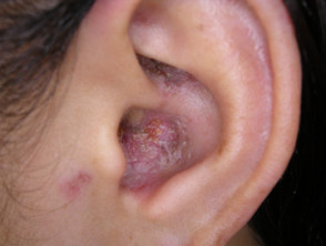
- DLE tends to appear in exposed areas such as the nose, cheeks, earlobe, or pinna.
- There is a possibility of involvement of the lips, the mucosa oral mucosa, nose, or eyelids.
- Lesions on the scalp cause patches of hair loss, which can be temporary or permanent.
- Hypertrophic (or verrucous) lupus erythematosus Lupus erythematosus. hypertrophic (or verrucousis distinguished by notably thickened red plaques.
The specific signs of generalized DLE include:
- Plaques located on the anterior chest region anterior, the upper back, and the back of the hands.
- In some instances, plaques appear on the upper and lower extremities.
- There is a possibility of involvement of the palms and soles of the feet.
- Anogenital mucosa involvement may occur. anogenital area.
Although the main concern for the patient lies in the unsightly appearance of the plaques, these lesions can also manifest with localized itching or pain sensations.
Diagnosis and Classification of Chronic Discoid Lupus Erythematosus (DLE)
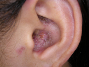
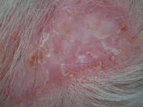
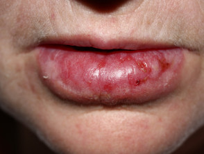
It is possible to consult additional visual material illustrating the different presentations of cutaneous lupus erythematosus.
Procedure for the Diagnosis of Discoid Lupus Erythematosus
DLE diagnosis is frequently established based on its typical distribution distribution in sun-exposed areas and confirmed by detailed clinical examination of the plaques. After conducting a thorough history, the patient suspected of having DLE should undergo a comprehensive physical examination to rule out the presence of any other lupus variant.
To establish a definitive diagnosis, a skin biopsy biopsy is generally required. This analysis will reveal the pathognomonic findings of lupus, including: interface dermatitis, periannexial involvement, follicular plugging, atrophy, and subsequent scar formation. Direct dermatitis interface dermatitis, periannexial involvement and, follicular plugging, atrophy, and the subsequent formation of scars. Direct
The immunofluorescence immunofluorescence frequently yields positive results in the lesional skin lesional in the direct study, confirming a positive lupus band test.
Cutaneous Lupus Erythematosus Disease Area and Severity Index (CLASI)
The Cutaneous Lupus Erythematosus Disease Area and Severity Index (CLASI) was developed as a systematic method to classify the severity of cutaneous LE. [2] This index calculates a score reflecting the activity and resulting damage from the disease, evaluating 12 defined anatomical locations (for all details, the original publication must be consulted).
The total activity score is calculated from the following parameters:
- Degree of erythema (score from 0 to 3) and scale (score from 0 to 2).
- Mucosal membrane involvement mucosa (score from 0 to 1).
- Recent hair loss (score from 0 to 1) and the presence of non-scarring alopecia alopecia (score from 0 to 3).
On the other hand, the total damage score includes:
- Level of depigmentation depigmentation (score from 0 to 2) and the degree of scarring (score from 0 to 2).
- Depigmentation persisting for more than 12 months doubles the score assigned to depigmentation.
- Specific score for scars present on the scalp (values: 0, 3, 4, 5, 6).
Blood Tests in Discoid Lupus Erythematosus
Patients with suspected or confirmed diagnosis of discoid lupus erythematosus (DLE) usually require complementary blood tests. These tests are essential to evaluate any possible systemic involvement and rule out other lupus variants, thus facilitating the design of a comprehensive management plan.
Generally, patients with discoid lupus erythematosus (DLE) will undergo blood draws both at the time of diagnosis and periodically thereafter:
- Although laboratory tests do not confirm the diagnosis, they are crucial for ruling out associated pathologies. Recommended investigations include:.
- Renal function tests.
- Measurement of inflammatory markers such as C-reactive protein Low (CRP).
- Antinuclear Antibody (ANA, ANF; if detected, they usually present in low titers).
- Extractable Nuclear Antibody (ENA).
- Anti-Annexin 1 Antibodies: These may function as a specific diagnostic marker for discoid cutaneous lupus erythematosus.
According to studies, circulating autoantibodies are detected in about 50% of individuals suffering from DLE.
Complications Associated with Discoid Lupus Erythematosus
A considerable proportion of patients, nearly 25% of those diagnosed with discoid lupus erythematosus, may later evolve into systemic lupus erythematosus. This transition can manifest months or even decades after the initial diagnosis of cutaneous involvement.
It is important to note that discoid lupus erythematosus has the potential to leave permanent scarring sequelae, even when the active phase of the disease has already responded satisfactorily to implemented therapies.
Treatment Protocols for Discoid Lupus Erythematosus
Fundamental Preventive Measures
Strict adherence to the following practices is essential to minimize the recurrence of DLE flares and control the disease:
- Establish a rigorous year-round sun protection routine. This involves the constant use of protective clothing, appropriate accessories, and the generous application of broad-spectrum sunscreens with a Sun Protection Factor (SPF) equal to or greater than 50+. It is crucial to understand that relying solely on sunscreens is not sufficient.
- In indoor environments, it is necessary for some patients to avoid direct exposure to solar radiation filtered through window glass, or to install protective films with UV blocking on them.
- Vitamin D supplementation is recommended for patients who present with deficiency.
- Patients who must strictly avoid exposure to solar radiation.
- Complete elimination of the smoking habit is fundamental.
Current Pharmacological Therapy for DLE
The fundamental treatment for Discoid Lupus Erythematosus (DLE) is based on the cyclical administration of high-potency topical corticosteroids corticosteroids . These must be applied precisely directly onto the affected skin areas for several consecutive weeks. The selection of the steroid potency must be done with extreme care, considering the body location and the specific thickness of the lesional plaque. The use of extremely potent topical steroids carries the potential risk of promoting thinning of the surrounding skin and accentuating the appearance of tiny blood vessels (telangiectasia). In specific situations, intralesional corticosteroid injections are considered, being especially effective for treating hypertrophic DLE.
Additionally, application of calcineurin inhibitors and such as tacrolimus ointment and pimecrolimus cream can be chosen.
The use of camouflage makeup is advisable to improve the aesthetic appearance of existing lesions.
Systemic Pharmacological Treatment
Generally, any of the following pharmacological agents can be implemented to treat DLE, either individually or in combination. It is crucial to highlight that the efficacy of these treatments decreases notably in patients who continue to smoke compared to those who abstain.
- Hydroxychloroquine and other antimalarials: documented response rates in cutaneous lupus (CL) are around 80%.
- Systemic corticosteroids, including prednisone or prednisolone. Their use for the exclusive treatment of DLE is infrequent.
- Methotrexate: Shows better tolerance and response in subacute cutaneous lupus and discoid lupus erythematosus.
- Corticosteroids Retinoids.
- Mycophenolate
- A biopsy may reveal deposits of
- Dapsone
- Thalidomide
- Belimumab
Prognosis of Discoid Lupus Erythematosus (DLE)
DLE tends to be a persistent condition persistent, lasting for significant periods that can extend for years or even decades. However, in a subset of patients, all parameters indicating disease activity resolve over time.
There is a rare possibility that a squamous cell carcinoma carcinoma of squamous cells may evolve within a long-standing DLE plaque, either on the skin or in a mucous membrane membrane pemphigoid. Clinically, this often presents as an actively growing, warty proliferation or as a cutaneous ulcer ulcer . A complication of this nature generally requires surgical intervention for proper management.


