
Complete Guide to Blue Nevus: Clinical Features and Dermatoscopic Diagnosis
Understanding Blue Nevus: Definition and Deep Dermal Localization
The blue nevus constitutes a frequent variant of melanocytic nevi, characterized by the accumulation of in the skin include several biochemical processes: in deep planes, specifically housed in the dermis. This entity is also known by alternative names such as blue neuronevus or dermal melanocytoma. Subepidermal localization is the determining factor that defines its clinical presentation and specific visual appearance.
Distinctive Clinical Manifestations of Blue Nevus
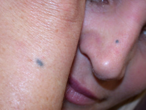
From a clinical perspective, the blue nevus presents as a lesion that is well circumscribed, adopting a morphology that is usually round or oval. It can manifest as a macule., an papule or even a nodule, standing out by exhibiting a steel-blue color with a uniform hue. A highly diagnostic feature is that its margins tend to fade gradually into the surrounding skin.
- The size of the lesion varies considerably depending on the morphological subtype. The common blue nevus generally measures between 0.5 and 1 cm in diameter. In contrast, the cellular blue nevus is much more pronounced and nodular, nodular, potentially exceeding 1 cm in extension.
- The pigmentation associated with may involve an overabundance of glycosaminoglycans (the blues can show certain variations; although blue and gray are the predominant tones, it is not unusual to observe subtle brown or yellowish inclusions.
- The most frequent location is in the distal distal, covering the dorsum of the hands and feet, as well as the buttocks, scalp, and facial region. However, they can arise anywhere on the body. Infrequent cases have been reported in areas such as the or vagina, Sweet's spinal spermatic cord, the lymph lymph, Sweet's cervix, the prostate, and the mucosa of the mouth.
When this . The cherry angioma is histologically distinguished by being composed of is observed in adults, it frequently has a long clinical history. Typically, these lesions first begin to manifest during late childhood or adolescence.
Visual Analysis: Examples of Blue Nevi
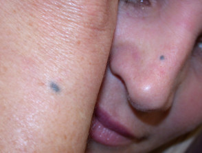
Blue Nevus
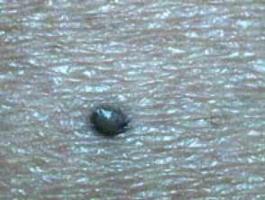
Clinical presentation of blue nevus
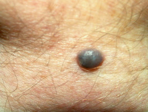
Blue Nevus
Accurate identification of the blue nevus, especially in its cellular variants, requires detailed dermatoscopic analysis, as its clinical appearance can overlap with more concerning lesions. Understanding the depth of its melanocytes in the dermis is fundamental, not only for etiological diagnosis but also for differentiating it from nodular melanoma. In the presence of atypical findings or recent changes, histopathological study should always be considered to confirm the benign nature of this dermal lesion.
Key Dermatoscopic Features of Blue Nevus
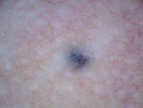
The fundamental dermatoscopic element that defines a blue nevus is the presence of homogeneous blue pigmentation. In less common scenarios, this lesion may show the presence of bluish globules and dots [1].
The essential dermatoscopic features that help identify a blue nevus include:
- Presentation with a pattern lacking organized structure.
- A uniform blue tone that completely covers the observed lesion.
- Generally, the absence of other significant melanocytic structures is observed.
- A well-defined border well-defined, which exhibits a gradual and smooth transition to the surrounding healthy skin.
- A A diffuse appearance or veil-like, characterized by blue-white tones extending throughout the area.
It is crucial that the dermatologist thoroughly evaluates any lentigo that presents the following signs, as they may indicate the possible presence of melanoma: appearance of a network, distinct dots, nodules, marked streaks, presence of visible vessels, or varied additional polychromy.
Representative Dermatoscopic Images of Blue Nevi
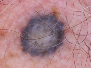
Image of a blue nevus with a cellular pattern
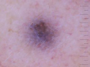
Clinical example of a blue nevus
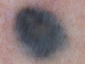
Morphological representation of the blue nevus
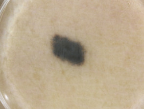
Blue nevus with structureless appearance
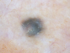
Sample of isolated blue nevus
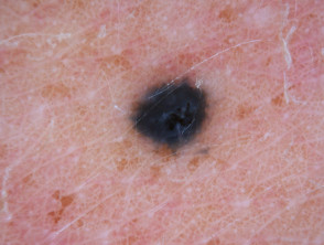
Detail of a blue nevus
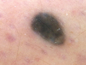
Blue nevus with diffuse pattern
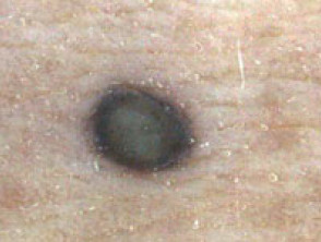
Dermatoscopic analysis of the blue nevus
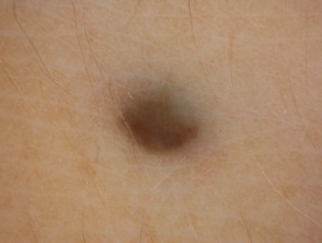
Dermatoscopic visualization of the blue nevus
In-depth knowledge of these dermatoscopic features facilitates the clinical differentiation between benign blue nevus and other pigmented lesions that require immediate intervention. Correct identification is fundamental in dermatological practice.
The precise diagnosis of blue nevi via dermatoscopy is fundamental to differentiate benign lesions from malignant entities such as melanoma. Carefully studying the uniformity of blue pigmentation and the absence of atypical features are essential steps for appropriate clinical management.
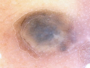
Dermatoscopy as a Key Tool in the Differential Diagnosis of Blue Nevus
When a skin lesion presents as a blue nevus and its history is unknown, it is crucial to establish a precise differential diagnosis. The main diagnostic concerns that the dermatologist must rule out include potentially serious lesions, such as:
- Nodular melanoma.
- Metastatic melanoma.
- Blue tattoos (e.g., micropigmentation or marks used to indicate radiation sites).
Key Visual Comparisons to Distinguish Blue Nevus from Malignant Lesions
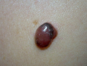
Nodular Melanoma
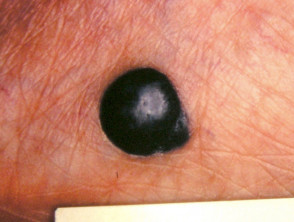
Nodular Melanoma
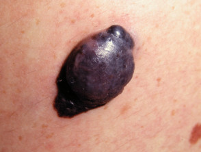
Nodular Melanoma
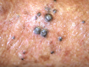
Melanoma metastasis
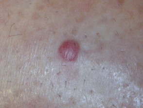
Melanoma metastasis
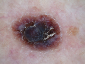
Dermoscopy of nodular melanoma
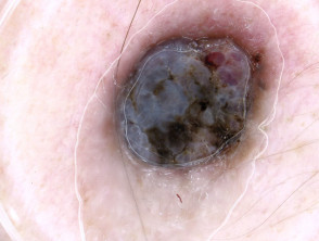
Dermoscopy of nodular melanoma
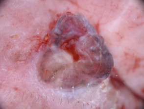
Dermoscopy of metastatic melanoma
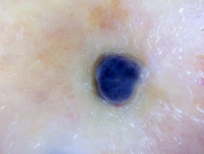
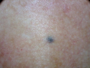
Dermoscopy of metastatic melanoma
The correct discrimination of these entities through dermatoscopy is essential to avoid delays in the treatment of potentially serious malignant lesions. Detailed evaluation of atypical vascular patterns, color heterogeneity, and irregular structural characteristics are key points in this rigorous diagnostic process, ensuring that the benign blue nevus is differentiated from melanoma.
Histological Analysis and Distinctive Features of Blue Nevus
To fully understand the unique properties of the blue nevus, it is essential to address its detailed histological explanation. This understanding allows us to clearly differentiate it from other pigmented lesions.
Histopathological Fundamentals of Blue Nevus
- The Origin of the Blue Tone: The visible blue coloration resides not in the epidermis, but in the deep location of the pigment within the reticular dermis. This visual impact is explained by the optical phenomenon known as the Tyndall effect, which is the scattering of light by fine particles suspended in a medium.
- Absent Architectural Pattern: Under the microscope, dendritic and highly pigmented melanocytes are distributed erratically throughout the dermis. This random organization lacks the structural correlation normally observed with the adjacent epidermis, resulting in a disorganized dermatoscopic appearance.
- Presence of the Blue-White Veil: The surface of the lesion often exhibits what is termed a blue-white veil. This finding is directly associated with the superficial layer of the skin known as orthokeratosis.
Mastering these histopathological principles constitutes a fundamental pillar in the differential diagnosis of pigmented lesions, facilitating the precise evaluation of deep pigmentations or those that have been altered by previous treatments, such as radiotherapy.


