
Dermoid Cyst: Histological Features and Differential Diagnosis
Dermoid cysts originate from the ectoderm and interact with the embryological closure lines of development. These benign tumors present distinctive histological features that are crucial for their pathological identification.
Histopathology of the Dermoid Cyst
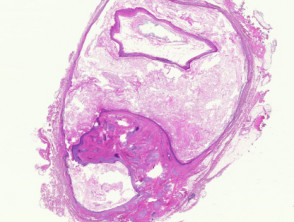
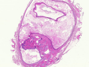
When examining histological sections of a dermoid cyst, a lesion is frequently observed subcutaneous that often appears «flaked off» or detached during processing (Figure 1).
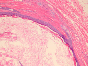
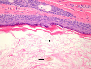
The characteristic inner lining is a keratinizing squamous epithelium (Figures 2, 3). The hallmark of these cysts is the presence of evident pilosebaceous appendages pilosebaceous integrated into the cystic wall (Figures 2, 3). Furthermore, hair material (vellus) is commonly found within the cyst lumen (Figure 3, arrow).
Less frequent but relevant findings in the cyst wall may include a mucinous or mucus-producing secretory epithelium, mucus, the presence of smooth muscle, and nodes sweat eccrine surrounding them.
Illustrated Pathology of the Dermoid Cyst
Specialized Studies for Dermoid Cyst
In general, routine histopathological studies are sufficient; therefore, no additional special studies are required to confirm the diagnosis of a dermoid cyst.
Differential Diagnosis of a Dermoid Cyst
It is crucial to distinguish the dermoid cyst from other cystic lesions, especially the steatocystoma. Steatocystoma typically exhibits sebaceous glands sebaceous directly connected to the cystic lining, but it lacks the pilar structures observed in the dermoid. Additionally, the lining of the steatocystoma does not present a granular layer and shows a characteristic wavy or "border" basal layer (kerb). layer granulomatous and shows a characteristic wavy or "border" basal layer (nail edge).


