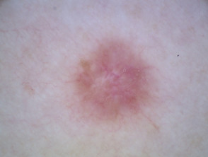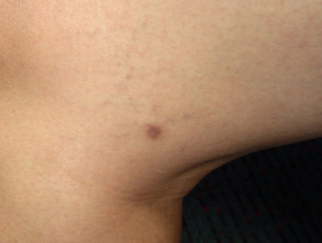
Images of Dermatofibroma

2402__protectwyjqcm90zwn0il0_focusfillwzi5ncwymjisingildfd-7014202-4764396-jpg-8645015

Table of Contents

«`html

Go to the image library

Visual Gallery: Examples of Classic Dermatofibroma
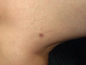
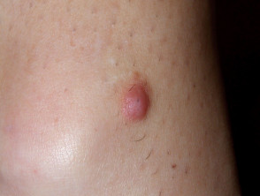
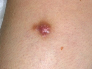
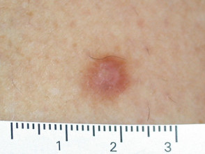
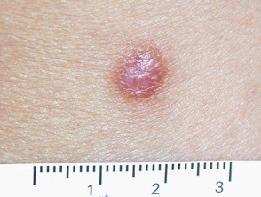
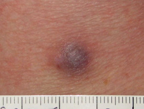
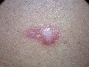
Typical morphology of a dermatofibroma on the skin.

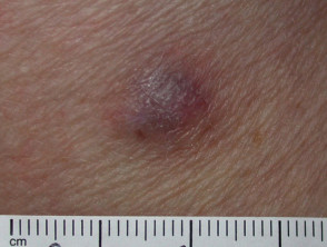
Skin lesion presenting an elevated dermatofibroma.

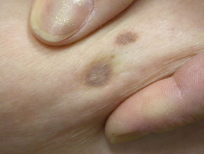
Clinical appearance of a dermatofibroma with varied coloration.

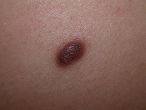
Dermatofibroma showing its well-defined contour.

Example of dermatofibroma in an initial or regressive phase.

Nodular dermal lesion characteristic of dermatofibroma.

Comparison of several dermatofibromas in the same patient.

Dermatofibroma located in a sun-exposed area.

Dermatofibroma: small, firm nodular lesion.

Dermatofibroma

Secondary image of a dermatofibroma.

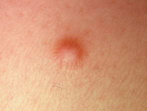
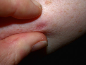
Positive pinch test in a dermatofibroma.

Pinch test

Characteristic central dimpling sign of a dermatofibroma.

Pinch test

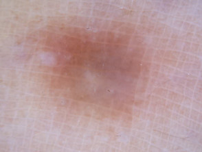
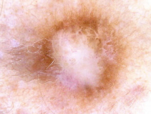
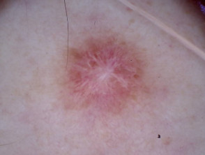
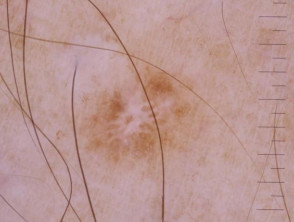
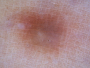
Dermoscopic Examination: Dermatofibroma Under the Microscope
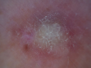
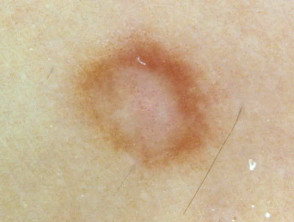
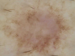
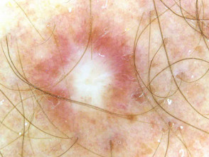
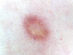
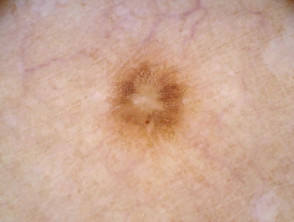
Dermoscopic view of a dermatofibroma highlighting vascular patterns.

Dermatofibroma

These images complement the clinical information regarding the diagnosis and visual characteristics of dermatofibroma, a very common benign skin lesion. Reviewing these examples helps in correctly identifying the lesion through physical examination and, when necessary, through dermoscopy.

«`
Dermatly.com - El sitio de tu piel