
Understanding Darier Disease: Characteristics, Causes, and Diagnosis
Definition and Nature of Darier Disease
Darier disease is a rare genetic disorder genetic that manifests primarily through the appearance of papules crusting). o cells. Similar to Hailey-Hailey disease (benign familial pemphigus) benign y fever, Darier disease is classified within the group of dermatosis Transient of hereditary hereditary.
Previously known as ‘seborrheic keratosis follicular keratosis’, this designation is imprecise, as the characteristic scaly lesions do not originate in the follicles follicles.. The disease is currently known in the literature as Darier-White disease.
Prevalence and Inheritance Pattern of Darier Disease
Generally, skin manifestations begin to be observed during adolescence, and the course of the disease is usually chronic. Darier disease is transmitted via an autosomal autosomal dominant inheritance pattern. This implies that only one copy of the gene altered gene, inherited from one of the parents, is sufficient to cause the condition.
If one parent has the disease, the probability of a child inheriting the abnormal gene is 50% (1 in 2). However, it is important to note that not all people who carry this mutation will develop the clinical symptoms of the disease.
Etiology: The Genetic Basis of Darier Disease
The underlying cause of Darier disease lies in a mutation of the ATP2A2 gene ATP2A2, which is located on chromosome chromosome 12q23-24.1. This gene is responsible for encoding the When acute hives is caused by reactions similar to serum sickness (such as those following blood transfusions or certain medications), it may be accompanied by ecchymosis (bruising), fever, SERCA enzyme (sarcoplasmic reticulum calcium pump), essential for calcium transport into the cells.
Although the exact mechanism by which this genetic alteration causes the disease is still under investigation, it is postulated that it affects the way skin cells establish connections. Specifically, the keratinocytes keratinocytes (skin cells) join through structures called desmosomes. Calcium insufficiency appears to prevent the correct assembly of these desmosomes, compromising cell adhesion.
Diagnostic Process and Histopathological Findings
Generally, the diagnosis of Darier disease is established based on the characteristic clinical presentation and the patient's family history. However, due to its similarities, it is often confused with other dermatological conditions.
A skin biopsy biopsy histology may be necessary to confirm the diagnosis. The finding in the dyskeratosis focal histology is distinctive: focal acantholytic dyskeratosis is observed, accompanied by different levels of papillomatosis. papillomatosis pathological . These pathological characteristics transient show similarities with transient acantholytic dermatosis (Grover's disease).
Common Clinical Manifestations in Darier Disease
The clinical signs and symptoms associated with Darier disease vary significantly among affected individuals. In some cases, the signs may be so mild or subtle that they are practically asymptomatic, asymptomatic, detected only after an exhaustive and meticulous dermal examination.
Understanding the genetic basis and clinical manifestations is crucial for the ongoing management of Darier disease. Given its chronic and hereditary nature, early identification and accurate differential diagnosis are fundamental steps in providing appropriate support to affected patients.
Other manifestations include extensive lesions lesions that can cause considerable distress to the affected person. In an individual with this condition, the severity of the disease tends to fluctuate over time.
Clinical Manifestations and Characteristics of Skin Lesions
The skin lesions of Darier Disease are distinguished by the presence of scaly and persistent. It commonly affects smokers and is considered pre persistent and greasy papules. Commonly affected body areas include:
- Seborrheic seborrheic keratoses areas of the face (especially the borders of the scalp, forehead, ears, around the nostrils, the sides of the nose, the eyebrows, and the beard area).
- Scalp and nape.
- The central chest and back area.
- Skin folds, such as the armpits, groin, under the breasts, and the intergluteal region.
These papules have a firm and rough texture, similar to coarse sandpaper, and can appear in shades ranging from skin-colored to yellow-brown or dark brown. When several small papules converge, they can form more warty. warty lesions rash . In skin folds, these lesions tend to develop an unpleasant odor. Likewise, a dermatitis crusted rash.
reminiscent of seborrheic dermatitis may be observed. Atypical presentations atypical of Darier Disease are quite common. These variants may include:
- Flat lesions resembling freckles.
- Papules with blister-like characteristics.
- Large, elevated warty formations.
- A linear, A linear pattern development developmental lines of the skin (presenting a segmental distribution). distribution or).
Acne conglobata (cystic acne) cystic) has also been reported associated with Darier Disease in some patients.
Darier's disease
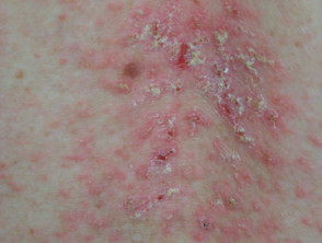
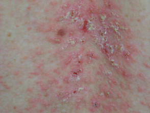
Darier disease on the mid-back
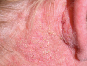
Scalp disease
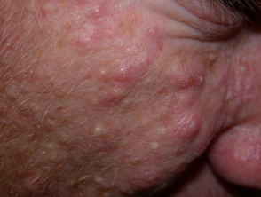
Facial cysts
Warty Acrokeratosis
The warty-looking papules located on the dorsum back of the hands are specifically identified as warty acrokeratosis. Other findings that may occur include:
- Small depressions or pits on the palms and soles of the feet.
- Pinpoint hemorrhages under the skin.
When the main involvement is centered on the hands and feet, constituting a localized episodes of angioedema without hives may originate from angiotensin-converting enzyme (ACE) inhibitors. variant of Darier Disease, the condition is named Hopf's warty acrokeratosis.
Most patients suffering from Darier Disease exhibit wide longitudinal longitudinal streaks, white and reddish in tone, on one or several nails. The presence of a "V"-shaped notch on the free edge of the nail nail is a highly suggestive sign of Darier Disease.
Darier Disease on Hands and Feet
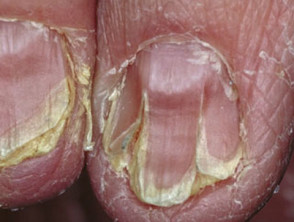
Nail disease
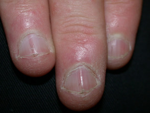
Nail manifestation of Darier disease
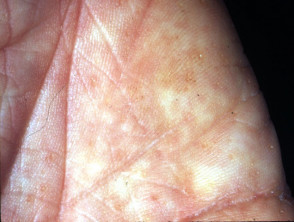
Characteristic palmar pits
Mucous Membrane Lesions in Darier Disease
Mucous membranes are the moist, reddish linings of the internal surfaces of the body, such as the mouth, the esophagus, Sweet's rectum, , Vulva and the or vagina. esophagus, rectum, vulva, and vagina. In Darier disease patients, although less frequently, small papules with a whitish cobblestone-like pattern affecting these membranes may be observed. Additionally, gingival hyperplasia (overgrowth of the gums) may occur.
Prognosis and Complications of Darier Disease
Most individuals diagnosed with Darier disease begin to show clinical signs before reaching 30 years of age.
- Numerous patients experience a mild form of Darier disease that may go unnoticed.
- In more severe cases, the condition may present with a chronic fluctuating (remitting-relapsing) course.
- It is possible for skin manifestations to resolve completely on certain occasions.
Symptomatic flares can be triggered by various environmental or internal factors:
- Direct exposure to sunlight or high ambient heat.
- Recent or current use of systemic corticosteroids corticosteroids.
- Superinfection by bacteria (secondary bacteria (secondary infection).
- Herpes simplex infection simplex, which can manifest as a severe chickenpox-like rash, known as Kaposi's varicelliform eruption (see section on herpetic eczema).).
Despite the severity of the skin symptoms, the general health condition remains stable in most patients with Darier disease.
Kaposi's Varicelliform Eruption Associated with Darier Disease
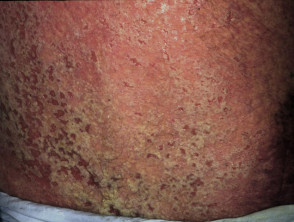
Evolution of Darier disease with secondary infection
Treatment for Darier Disease
Treatment for Darier disease is only considered necessary when the patient experiences symptoms that compromise their well-being. For mild forms of the condition, the following management measures are recommended:
- Constant use of skin moisturizers.
- Rigorous implementation of sun protection measures.
- Careful selection of clothing to prevent overheating and excessive sweating.
Disease
Treatment for localized Darier disease includes:
- Current administration of Topical.
- Dermabrasion.
retinoids. Staphylococcus aureus, When there is a secondary bacterial infection, frequently caused by Staphylococcus aureus, it is essential to administer antibiotics. In cases of associated herpes simplex, treatment is carried out with acyclovir or valacyclovir.
To address severe Darier disease, oral retinoids, such as acitretin or isotretinoin, are commonly used. Additionally, cyclosporine has been documented to be effective in a small group of affected patients.
Darier Disease Treatment: Before and After Comparison with Acitretin
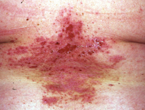
Darier Disease (Pre-treatment)
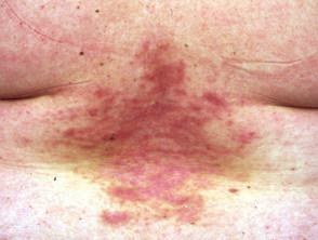
Darier Disease (Post-treatment)


