
These rare cysts probably originate from various developmental anomalies, depending on their anatomical location. The most frequently documented variant is the «cutaneous ciliated cyst of the lower limb.» This type appears almost exclusively on the lower extremities of women.
Histology of the Cutaneous Ciliated Cyst
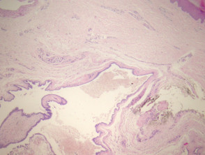
In the cutaneous ciliated cyst, histological sections reveal a dermal-based cyst (Figure 1) that is frequently filled with secretions and cellular debris (Figure 2). The cyst wall is lined by a epithelium cuboidal or columnar epithelium that exhibits cilia (Figures 3 and 4; arrows point to the cilia). Areas of squamous metaplasia (metaplasia) may be observed. squamous metaplasia (squamous metaplasia).
Pathology of the Cutaneous Ciliated Cyst
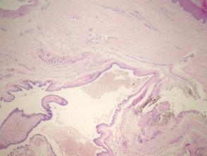
Figure 1
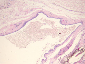
Figure 2
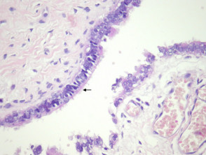
Figure 4
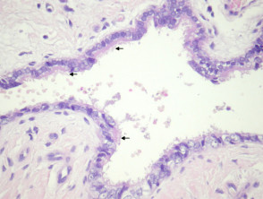
Figure 5
Special Studies for the Cutaneous Ciliated Cyst
Generally, no special ancillary studies are required for this diagnosis.
Differential Diagnosis of Cutaneous Ciliated Cyst Pathology
Bronchogenic Cyst: Usually presents smooth muscle, nodes and occasionally, cartilage in its wall. These cysts usually manifest in the midline, midline, near the sternum.
Branchial Cleft Cyst: Typically possesses a squamous lining, although it may exhibit foci of glandular epithelium glandular fever) with the presence of cilia. The wall frequently contains lymphoid tissue lymphoid. Smooth muscle and cartilage are also commonly observed in the structure of its wall.


