
Histopathological Diagnosis of Parapoxvirus: Milker's Nodule
The nodule Milker's nodule (caused by Parapoxvirus in cows), bovine papular Papular stomatitis (affecting the mucosa oral mucosa of cattle), and Orf (seen in sheep and goats) are viral infections that have zoonotic potential, capable of being transmitted to humans.
Distinctive Histological Features of Milker's Nodule
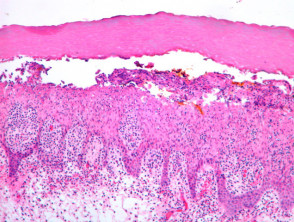
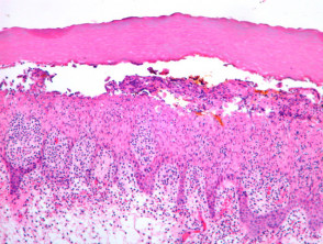
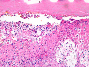
When examining a papulovesicle typical and well-developed of milker's nodule, histological analysis reveals an intraepidermal blister. intraepidermal. This lesion originates from massive swelling of infected keratinocytes, followed by marked necrosis cell necrosis (Figure 1). Acute spongiosis is commonly seen spongiosis acute along with this necrotic process.
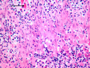
Intranuclear inclusions, which stain with an eosinophilic color, eosinophilic, are often very subtle and difficult to precisely locate (Figure 3, arrow). Frequently, the histopathological diagnosis relies on the predominant tissue reaction pattern, characterized by necrosis and degeneration of the keratinocytes edematous keratinocytes. A clinical history involving exposure to farm animals is essential, sometimes requiring complementary studies to confirm the exact etiology.
Visualization of Milker's Nodule Pathology
Techniques for the Specific Diagnosis of Milker's Nodule
For the specific visualization of the characteristic inclusion bodies inclusion associated with the milker's nodule virus, the application of Lendrum stain using phloxine-tartrazine is useful, which highlights these structures more clearly.
In addition to morphological analysis, direct confirmation of viral presence can be achieved through cell culture technique, the culture cell test, the antinuclear antibody fluorescent antibody (FTA) test, or using advanced techniques such as electron microscopy. microscopy microscopy.
Differential Diagnosis in Milker's Nodule Pathology
The pathology Milker's nodule pathology presents overlapping characteristics with other infectious diseases, including vaccinia, equine pox, and human smallpox. All these pathologies share prominent keratinocytic swelling, necrosis, and the presence of inclusion bodies that can be morphologically similar to each other.
In these confusing diagnostic scenarios, a thorough clinical history and, if necessary, additional microbiological studies, are indispensable to ensure an accurate final diagnosis. In contrast, infections caused by herpes simplex virus (HSV), as well as chickenpox and herpes zoster, present viral inclusion bodies with different structural architectures that facilitate their distinction. simplex herpes simplex virus (HSV), as well as chickenpox and herpes zoster, present viral inclusion bodies with different structural architectures that facilitate their distinction.
The accurate diagnosis of milker's nodule, based on histopathology and epidemiological context, is a crucial step for the proper management of any suspected case in veterinary or human dermatology clinics.


