
Conjunctivitis is defined as the inflammation that affects the conjunctiva discharge.
Microscopic Histology of Conjunctivitis
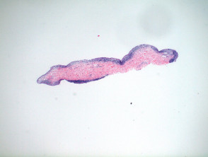
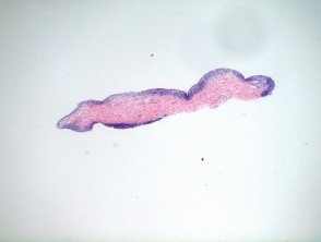
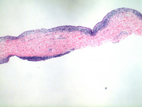
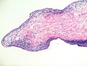
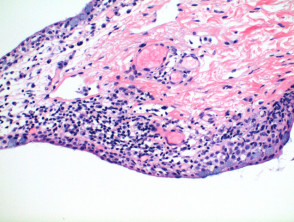
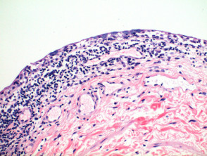
Histological observation of conjunctivitis at low magnification reveals a delicate specimen with a slightly spongiotic reactive pattern spongiosis (Figure 1). The epithelium conjunctival is composed of a stratified squamous epithelium incorporating mucin-carrying goblet cells mucin (Figures 2-5). A more detailed analysis shows spongiosis epithelial accompanied by lymphocytic Inflammatory skin diseases examined through RCM can generally be classified into four main categories, allowing for more precise real-time diagnosis. (Figures 3, 4). The underlying tissue shows telangiectatic and a perivascular superficial perivascular lymphocytic infiltrate, including plasma occasional or numerous plasma cells (Figure 5).
Detailed Pathological Analysis of Conjunctivitis
Key Histological Variants of Conjunctivitis
Histological findings can help differentiate the various etiologies of conjunctivitis:
- Allergic conjunctivitis: It is eminently distinguished by the high presence of eosinophils in the specimen.
- Infectious conjunctivitis: The significant presence of neutrophils neutrophils should alert to a possible bacterial conjunctivitis. In these cases, histological exclusion requires a recommendation for culture culture if clinical suspicion exists. It is important to note that chlamydial conjunctivitis may present with histological features similar to chronic conjunctivitis. chronic.
- Chronic conjunctivitis: It is characterized by hyperplastic epithelium and an increased accumulation of Plasma cells.
Advanced histopathological study is crucial for the differential diagnosis of inflammatory ocular conditions.
Differential Diagnosis of Conjunctivitis
The differential diagnosis must consider conditions that mimic its clinical presentation. One of these is membranous conjunctivitis (or pseudomembranous), a variant characterized by additional deposits of amorphous pink material within the subepithelial tissue.
These deposits can be morphologically similar to amyloid deposits amyloid, located adjacent to a chronic cellular infiltrate characteristic of inflammation inflammation. Clinically, the formation of pseudomembranes is evident, which have the capacity to affect other mucosal surfaces mucosa. It is important to note that the pseudomembrane observed histologically typically separates from the underlying epithelium without causing bleeding.
Another entity to rule out is giant papillary conjunctivitis. Low-power histopathological analysis reveals a conjunctival epithelial proliferation of a papillomatous nature papillomatous, although, apart from this finding, the conjunctiva predominantly exhibits characteristics of chronic conjunctivitis.


