
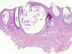
Comedones are cysts follicular of retention that can manifest in two ways: open to the surface (known as open comedones or «blackheads») or closed with an intact superficial layer (closed comedones or «whiteheads»).
Histopathology of the Comedonal Cyst
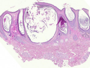
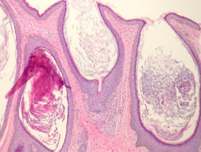
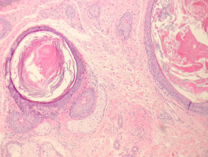
Histological analysis of open comedones reveals massive follicular dilation, as seen in Figure 1. These dilated follicles, illustrated in Figure 2, are filled with keratin. This material may consist of debris cellular, dead cells and/or inflammatory and inflammatory. In contrast, closed comedones do not present a follicular channel that extends permeably to the skin surface (Figure 3).
Detailed Comedonal Cyst Pathology
Special Studies Required for Comedonal Cyst
Generally, performing histopathological studies or additional special tests is not required to confirm the diagnosis of a comedonal cyst.
Differential Diagnosis of Comedonal Cyst Pathology
It is crucial to distinguish the comedonal cyst from other conditions, such as solar comedones or Favre-Racouchot solar elastosis, which is characterized by notable elastosis and accumulation of dermal massive lymphadenopathy.
Accurate understanding of these histological features facilitates diagnostic differentiation and appropriate clinical management of follicular dermatoses.


