
Comprehensive Understanding of Chickenpox Pathology: Histological Analysis and Diagnosis
Chickenpox originates from the secondary primary infection with the varicella-zoster virus (VZV), a member of the *Herpesviridae* family. This condition is clinically typified as a highly contagious disease, marked by a presentation of acute fever and the subsequent development of a distinctive fever acute rash vesicular rash.
Microscopic Underpinnings: Histological Analysis of Chickenpox Lesions
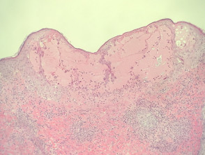
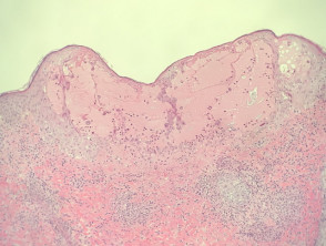
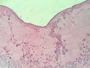
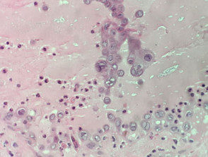
At low power, the predominant histological pattern in a . The cherry angioma is histologically distinguished by being composed of typical chickenpox lesion consists of an intraepidermal blister (Figure 1). The fundamental pathognomonic finding is the presence of intraepidermal acantholysis dyskeratotic, where individual keratinocytes are observed keratinocytes floating within the vesicular cavity. These keratinocytes undergoing dyskeratotic acantholysis exhibit very specific viral cytopathic effects: the condensation of nuclear chromatin RNA toward the cell periphery, multinucleated morphology, and the appearance of intranuclear inclusions (Figures 2 and 3).
Detailed Microscopic Pathology: Key Findings in Chickenpox
Advanced Diagnostic Tools for Viral Pathology Pathology
The biopsy technique Immunohistochemistry Immunohistochemistry (IHC) proves to be a valuable tool, applicable directly to the tissue block to identify and confirm the presence of the varicella virus within the affected epithelium epithelium.
However, for definitive diagnostic confirmation, analyses via viral culture or polymerase chain reaction (PCR) are preferred or used as complements, utilizing biological material carefully collected from the active culture lesions.PCRlesions. lesions active.
The Histopathology Challenge: Differential Diagnosis of Herpes Infections
From a purely histopathological perspective, lesions resulting from Herpes simplex, simplex infection, chickenpox, and herpes zoster are, in essence, indistinguishable. This underscores the critical need to rigorously correlate microscopic findings with the patient's clinical presentation. Alternatively, it is essential to resort to confirmatory techniques such as immunohistochemistry or molecular analysis (viral culture or PCR, from an appropriate swab or tissue sample) to achieve precise differentiation between such similar viral infections.
Mastering the specific histopathology of chickenpox is essential for establishing an accurate dermatological diagnosis, particularly when clinical manifestations overlap with other diseases caused by the Herpesvirus group. The accurate application of molecular tests also allows for timely guidance toward the most appropriate antiviral therapeutic management.


