
Calciphylaxis: Histopathological Features and Diagnostic Guide
Calciphylaxis predominantly occurs in patients suffering from end-stage renal disease (ESKD). This severe systemic disorder is primarily characterized by progressive **calcification** of blood vessels and surrounding soft tissue, which inevitably triggers cutaneous infarcts and extensive tissue necrosis.
Key Histopathological Findings in Calciphylaxis
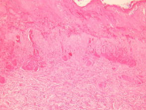
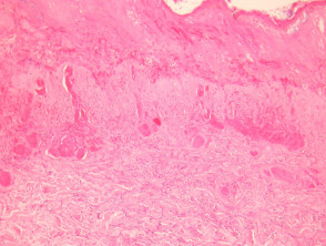
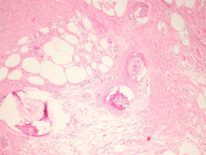
To establish a definitive diagnosis of calciphylaxis, it is imperative to obtain a deep **biopsy** that explores the subcutaneous tissue. Generally, the superficial **epidermis** and **dermis** show advanced ulcers and necrosis, along with multiple secondary reactive changes (Figure 1). The pathognomonic indicator is calcification affecting small and medium-sized distribution vessels (Figures 2 and 3).
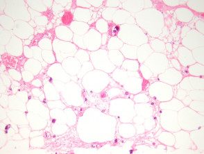
At the histological level, it is common to observe fibrosis in the intima layer of the vessels, and **intravascular thrombi** are frequently identified. A crucial defining sign is the **extensive** and diffuse **calcification** observed in the small **capillaries** embedded in the adipose tissue (Figure 3). Furthermore, secondary calcification of **adipocytes** may manifest.
Pathological Evidence of Calciphylaxis
Specialized Techniques for Diagnostic Confirmation
Unequivocal identification of calcium deposits is significantly facilitated by the use of specific stains. Von Kossa stain is the preferred method to visualize the presence of calcium in the obtained tissue samples.
Establishing the Differential Diagnosis with Other Conditions
An exhaustive anamnesis collection, highlighting a history of insufficiency chronic kidney and the appearance of lesions necrotic skin lesions, establishes a high clinical suspicion of calciphylaxis. This initial approach is fundamental.
It is crucial to differentiate this pathology from other entities that present with vascular calcification, such as atherosclerosis or diabetic complications. Although these conditions also involve some degree of calcification, the combination of vascular thrombosis and diffuse microvascular involvement of capillaries and fatty tissue is highly indicative of calciphylaxis, consolidating its diagnosis.


