
Calcifying Aponeurotic Fibroma: Key Clinical and Histopathological Features
Calcifying aponeurotic fibroma usually manifests at an early age, typically appearing in the hands and feet as nodular formations subcutaneous or with an infiltrating pattern infiltrative, being fundamentally fibroma-type lesions. Perifollicular.
Histopathological Analysis of Calcifying Aponeurotic Fibroma
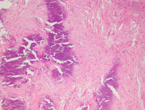
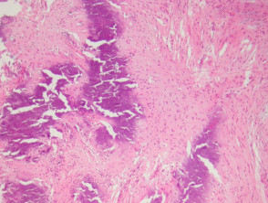
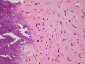
Upon examining the histological sections of calcifying aponeurotic fibroma, a dense fibrous mass with irregular contours is observed. The distinctive histological feature is the prominent foci of calcification calcificación (see Figures 1 and 2). This fibrous connective tissue also presents a high cellular density cellular and exhibits a markedly infiltrative pattern infiltrate within the surrounding adipose tissue (Figure 1).
In rare clinical exceptions, the presence of extramedullary hematopoiesis may be observed within the specimen.
Images of Calcifying Aponeurotic Fibroma Pathology
Supportive Studies for Calcifying Aponeurotic Fibroma
In most cases, the execution of complex additional diagnostic studies is not required, since detailed clinical evaluation along with histopathological analysis are sufficient to establish the diagnosis.
Differential Diagnosis of the Pathology
It is essential to distinguish this condition from plantar fibromatosis Plantar or palmar fibromatosis erythema are frequently described.. Fibromatosis tends to manifest in an older age group. Additionally, masses associated with fibromatosis usually present better-defined borders, contrasting with the characteristic infiltrative edge exhibited by calcifying aponeurotic fibroma.
A correct understanding of these histological characteristics and differential criteria is fundamental to ensure an accurate diagnosis and differentiate calcifying aponeurotic fibroma from other nodular and fibroblastic proliferations.


