
«`html
Essential Pathology and Histology of the Bronchogenic Cyst
Bronchogenic cysts originate from the ventral foregut, the embryonic precursor structure of the respiratory system. It is common to locate these pathological formations near the trachea or the main bronchi of the respiratory system.
Detailed Histological Analysis of the Bronchogenic Cyst
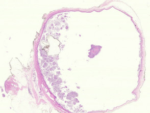
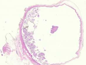
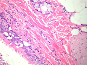
From a structural perspective, bronchogenic cysts can manifest as unilocular lesions or, alternatively, present a multilocular morphology. Although their appearance is less frequent in the skin, they can be found embedded in the dermis or subcutaneous tissue; occasionally, drainage toward the upper epidermis via a sinus is observed. Frequently, the interior of these cysts is obstructed with cellular debris and abundant thick mucinous material (Figure 2).
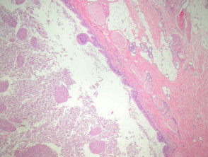
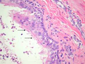
The internal lining of the cyst characteristically presents ciliated epithelium, which can be columnar or cuboidal (Figure 3, indicated by arrows). Squamous metaplasia is a common finding in this lining epithelium. Additionally, the cystic wall often includes smooth muscle and mucous glands that exhibit histological features typical of respiratory tissue (Figure 4). The presence of cartilage is another distinctive histological component frequently identified in the wall of these cystic lesions.
Key Pathological Manifestations of the Bronchogenic Cyst
Diagnostic Evaluation: Special Studies for Bronchogenic Cysts
Generally, the diagnosis of a bronchogenic cyst is established without the need for advanced or specialized complementary studies, based on histopathology.
Differential Diagnosis of Bronchogenic Cyst Pathology
Cutaneous Ciliated Cyst: The primary distinction is that ciliated cysts of cutaneous origin lack characteristic elements such as smooth muscle, mucous glands, and cartilage in their walls.
Branchial Cleft Cyst: This type of cyst typically exhibits a lining composed mostly of squamous tissue. However, it is important to note that it may present foci of glandular epithelium, some of which might contain cilia. It is common to observe lymphoid tissue present in the wall of a branchial cyst.
To confirm the histopathological diagnosis of the bronchogenic cyst and differentiate it from these similar entities, it is crucial to evaluate the presence of all typical respiratory components, such as ciliated epithelium, smooth muscle, and cartilage within the cystic matrix.
```


