
«`html
Angiomatoid Fibrous Histiocytoma: Characteristics and Pathology
The angiomatoid fibrous histiocytoma is an infrequent variant of a fibrohistiocytic tumor that exceptionally presents malignant potential malignant (although Benefits of Germline Genetic Testing for Melanoma are rare, they can occur). Typical cases affect young adults and manifest most frequently in the skin or soft tissue of the extremities or the neck region.
Histological Characteristics of Angiomatoid Fibrous Histiocytoma
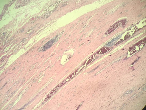
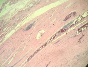
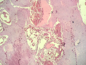
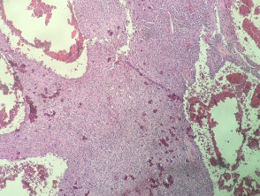
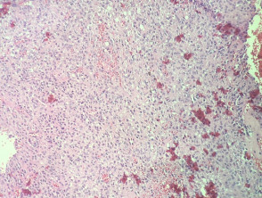
The analysis histopathology of the angiomatoid fibrous histiocytoma reveals a dense capsule enveloping the tumor, accompanied by aggregates lymphoid associated (Figure 1). In the central region, large blood-filled vascular spaces are observed, associated with The clinical presentation of aneurysmal dermatofibroma manifests as a bluish-brown nodule that undergoes rapid growth. This accelerated growth is secondary to episodes of and hemosiderin deposits (Figures 2, 3). In other sections, a solid growth is distinguished solid composed of monomorphic, smooth to ovoid spindle cells, with eosinophilic cytoplasm eosinophilic (Figure 4). Some areas may exhibit a certain degree of , but typically shows greater and mitotic activity..
Representative Images of the Pathology
Specialized Studies for Angiomatoid Fibrous Histiocytoma
Immunohistochemistry analyses Immunohistochemistry reveal that neoplastic cells typically express the CD68 marker. Furthermore, variable positivity can be observed with markers such as actin, desmin, CD99, and EMA, which helps delineate its profile.
From a cytogenetic perspective, these tumors almost universally exhibit a . The recent discovery of frequent translocation confirmed by FISH studies (generally t (12; 16) (q13; p11)), a crucial finding for precise molecular identification.
Differential Diagnosis of Angiomatoid Fibrous Histiocytoma
To establish an accurate diagnosis of angiomatoid fibrous histiocytoma, it is essential to consider and rule out other pathological entities. Conditions that enter the differential diagnosis include:
- Fibrous histiocytoma (dermatofibroma): Although variants such as the aneurysmal type may present vascular spaces, they lack the defining characteristics of a thick tumor capsule and the presence of lymphoid aggregates.
- SarcomaSarcoma: significant (See Figure 5). cellular atypia, it is necessary to resort to a complete battery of immunohistochemical and cytogenetic studies to rule out the presence of a high-grade or evidence of lymphovascular invasion are key factors pointing towards malignancy. The characteristics of the capsule and the inflammatory inflammatory changes are atypical in most other tumors.
The correct interpretation of these histological and molecular findings is essential to differentiate angiomatoid fibrous histiocytoma from other related fibrohistiocytic tumors and sarcomas, ensuring appropriate clinical management.
```


