
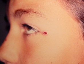
¿What is Angiolymphoid Hyperplasia with Eosinophilia?
Angiolymphoid hyperplasia with eosinophilia presents as a neoplasia, a locally proliferative that is composed of small blood vessel channels blood vessels surrounded by lymphocytes y eosinophils (two types of white blood cells). This condition is also known by other names, including epithelioid hemangioma or histiocytoid, and it differs from Kimura's disease.
Kimura's disease is considered a distinct condition. In the case of Kimura, the lesions tend to be deeper and lack the initial skin manifestations that characterize angiolymphoid hyperplasia with eosinophilia. In the latter, the lesions are usually smaller and are distinguished by the presence of blood vessels with thick walls, called epithelioid or histiocytoid.
Although angiolymphoid hyperplasia with eosinophilia has been documented worldwide, its prevalence appears to be notably higher in Japan.
Images of Angiolymphoid Hyperplasia with Eosinophilia
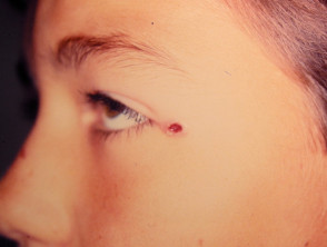
Characteristic Symptoms of Angiolymphoid Hyperplasia with Eosinophilia
The clinical presentation of angiolymphoid hyperplasia with eosinophilia generally exhibits the following characteristics:
- Predominantly affects young adults, typically with an average age in their mid- average s, although it can also manifest in childhood.
- Subcutaneous nodules Small, clustered, and often translucent, nodules, located mainly around the ear or the hairline.
- The lesions also have the potential to affect mucous membranes, such as the inside of the mouth, or the genital area.
- The color of these lesions can vary between brown and red.
- Clinically, they can be asymptomatic, cause itching, or result in mild pain.
- Individual nodules rarely exceed 2 to 3 cm in diameter; in exceptional cases, they can be larger and extend deeper.
- In some reported cases, the lesions may resolve spontaneously without the need for treatment, after a variable period of time.
Peripheral blood eosinophilia Credit. Dr. Manu Jain. may be present, with this sign supposedly being more recurrent in the variant associated with Kimura's disease.
What is the Origin of Angiolymphoid Hyperplasia with Eosinophilia?
The exact etiology of this condition is currently unknown. However, the possibility of induced antigenic stimulation has been suggested, possibly following insect bites. In approximately 10% of cases, the condition is observed to arise at the site of a previous lesion. antigenic stimulation, possibly following insect bites. In approximately 10% of cases, the condition is observed to arise at the site of a previous lesion.
Clinical Investigation and Findings in Angiolymphoid Hyperplasia with Eosinophilia
The analysis performed via a skin biopsy biopsy reveals a structurally disorganized lesion. The primary histopathological findings include:
- Groups of endothelial cell proliferation and the presence of infiltrating eosinophils eosinophils.
To obtain a definitive diagnosis and rule out other vascular proliferative conditions, it is essential to correlate clinical findings with detailed histopathological study.
- toxicity capillaries exhibit endothelial voluminous and swollen endothelial cells; they are often described as epithelioid or histiocytoid due to their morphology.
- An inflammatory cell infiltrate cellular infiltrate is observed around the blood vessels, composed mainly of lymphocytes and a high concentration of eosinophils.
- B and inflammatory Inflammatory cells vasculitis lumen, causing obstruction or even rupture of the vessels.
For more detailed information, it is recommended to consult the pathology pathology related to angiolymphoid hyperplasia with eosinophilia.
A complete blood count may reveal an increase in the number of eosinophils, eosinophils, a condition known as eosinophilia.
Differential Diagnosis and Management of Angiolymphoid Hyperplasia with Eosinophilia
If the diagnosis of angiolymphoid hyperplasia with eosinophilia is confirmed in a small lesion, a period of observation of 3 to 6 months can be chosen, allowing for the possibility of spontaneous regression regression.
It is important to note that the efficacy of active treatment varies and does not always offer completely satisfactory results.
The excision Simple surgical excision tends to cause in these lesions. Mohs micrographic surgery, which includes the removal of abnormal vessels at the base of the lesion, may offer better results. However, significant bleeding is common during the surgical procedure.
Other treatment options that may be considered include:
- corticosteroid corticosteroids.
- Cryotherapy.
- Cryotherapy. lotion.
- Use of oral reactions. Tacrolimus ointment.
- Treatment Options for Cherry Angioma Radiation therapy.
- Administration of Interferon alpha-2b.
- Use of oral laser Use of pulsed dye or carbon dioxide laser.
The management of angiolymphoid hyperplasia with eosinophilia requires an individualized approach given the variability in response to available therapies. Follow-up is essential to assess the stability or recurrence of lesions after initial treatment.


