
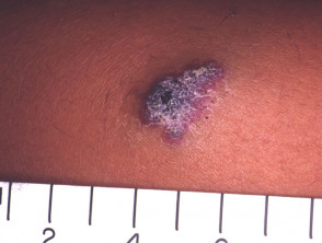
Images of Solitary Angiokeratoma
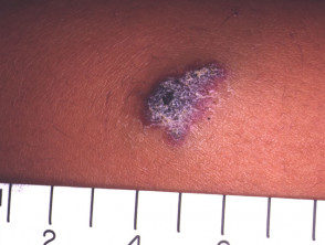
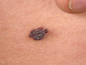
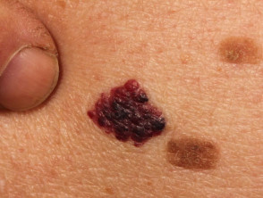
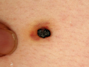
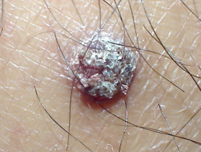
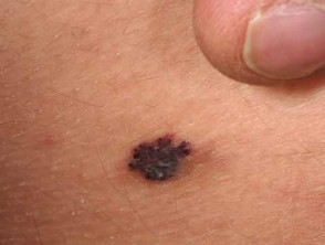
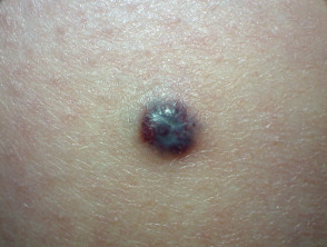
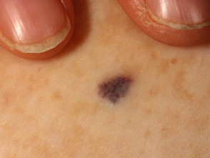
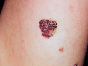
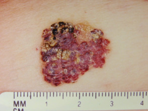
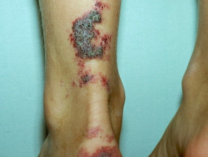
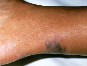
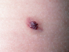
This gallery visualizes the different morphological presentations of Angiokeratoma, facilitating its recognition in dermatological differential diagnosis.
Fordyce Angiokeratoma: Characteristics and Clinical Presentation
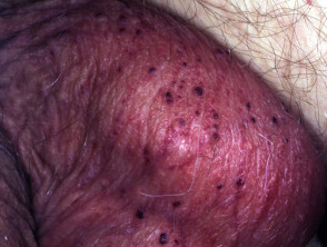
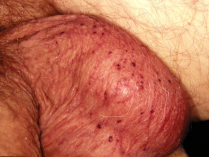
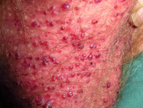
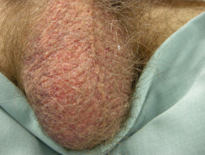
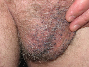
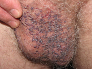
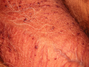
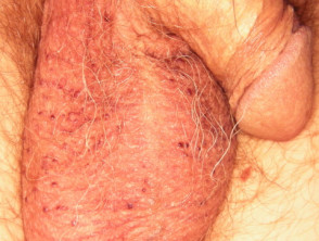
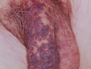
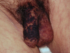
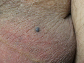
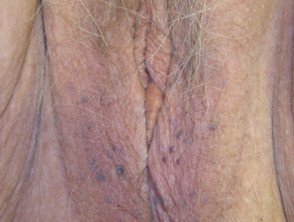
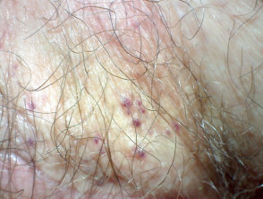
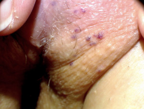
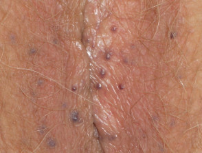

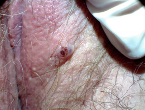
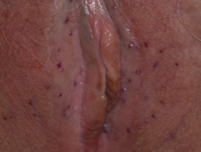
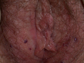
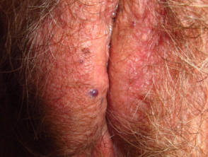
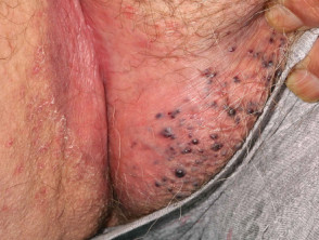
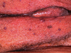
The provided content appears to be an image gallery without introductory text or analysis. Below, this content will be structured as a technical gallery, assuming the main topic is the visual manifestations of Angiokeratoma, focusing on Fordyce Angiokeratoma in the vulva, to provide appropriate SEO context.
Detailed Clinical Gallery: Manifestations of Angiokeratoma
Exploring the visual presentations of skin lesions is crucial for dermatological diagnosis. Below is a collection of images illustrating various aspects of angiokeratoma, with a particular emphasis on Fordyce's angiokeratoma, a benign vascular condition that frequently affects the vulvar region.
Angiokeratomas: Types and Visual Characteristics
Angiokeratomas are vascular malformations characterized by proliferation of dilated capillaries in the superficial dermis, covered by epidermal hyperkeratosis. While multiple types exist, the attached images focus on those requiring precise clinical differentiation.
Key Diagnostic Considerations
The correct identification of these lesions is fundamental, as angiokeratomas (especially Fordyce's) can simulate more serious lesions, such as melanomas or malignant vascular neoplasms. Dermoscopy light, as seen in one of the visual references, reveals distinctive reddish-black vascular dots and structures, helping the specialist confirm the diagnosis of Angiokeratoma without always resorting to invasive procedures.
Although these lesions are generally benign and asymptomatic, their location in sensitive areas or their cosmetically bothersome appearance may lead patients to seek treatment. The exhaustive review of these clinical images underscores the importance of continuous visual training in dermatology to adequately differentiate these vascular papules.
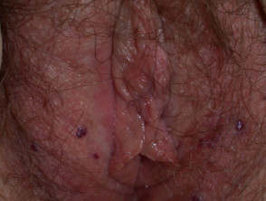
Dermoscopic Evaluation of Angiokeratoma
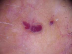
Dermoscopy of Fordyce angiokeratoma on the vulva
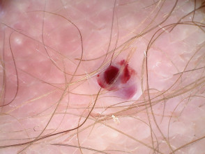
Dermoscopy of Fordyce angiokeratoma on the vulva
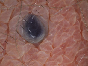
Dermoscopy of Fordyce angiokeratoma on the vulva
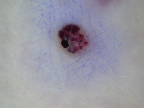
Dermoscopy of Fordyce angiokeratoma on the vulva
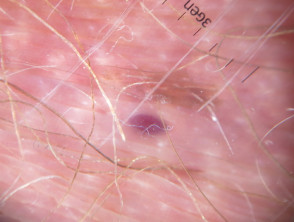
Dermoscopy of Fordyce angiokeratoma on the vulva
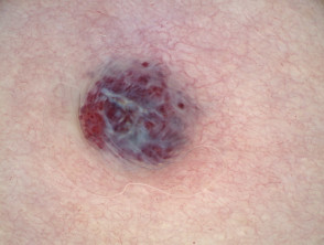
Dermoscopy of Fordyce angiokeratoma on the vulva
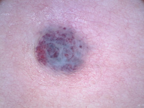
Dermoscopy of Fordyce angiokeratoma on the vulva
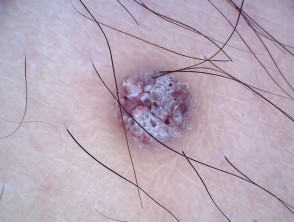
Dermoscopy of Fordyce angiokeratoma on the vulva
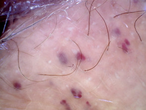
Dermoscopy of Fordyce angiokeratoma on the vulva
The precise diagnosis of dark or reddish skin lesions, such as angiokeratomas, often requires the use of dermoscopy. This non-invasive procedure allows the specialist to visualize subepidermal structures that are not evident to the naked eye, confirming key vascular characteristics associated with this condition.
Detailed inspection via dermoscopy is essential for differentiating angiokeratomas from other benign or malignant lesions, especially when located in sensitive areas like the vulva (Fordyce Angiokeratoma). Observing specific vascular patterns, coloration, and morphology helps establish an appropriate management plan.
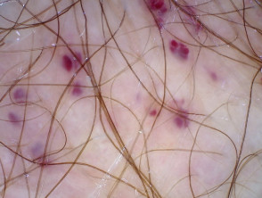
Dermoscopy of Fordyce angiokeratoma on the vulva
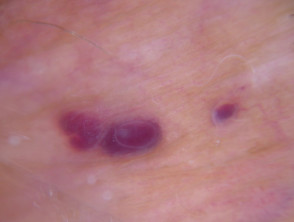
Dermoscopy of Fordyce angiokeratoma on the vulva
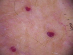
Dermoscopy of Fordyce angiokeratoma on the vulva
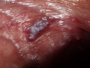
Fordyce's angiokeratoma on the vulva
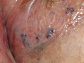
Fordyce's angiokeratoma on the vulva
Fabry Angiokeratoma: Cutaneous Manifestations
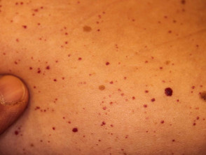
Angiokeratomas in suspected Fabry disease
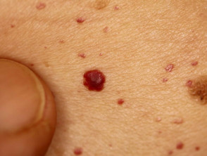
Angiokeratomas in suspected Fabry disease
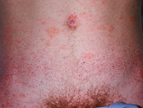
Angiokeratomas in Fabry disease
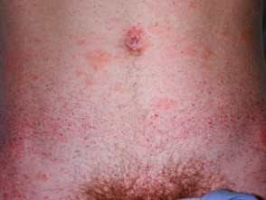
Angiokeratoma
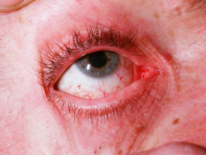
Angiokeratoma
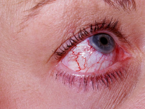
Angiokeratoma
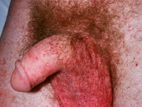
Angiokeratoma


