
Adenoid Cystic Carcinoma: Histological Features and Diagnosis
Adenoid cystic carcinoma generally originates in the nodes salivary. However, rare but recognized variants exist, such as primary cutaneous adenoid cystic carcinoma primary and breast adenoid cystic carcinoma.
Histology of Adenoid Cystic Carcinoma
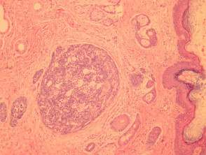
In primary cutaneous adenoid cystic carcinoma, primary, Sweet's tumor involves the dermis and has the capacity to invade subcutaneous tissue subcutaneously (Figure 1). Morphologically, the neoplasm is composed of basaloid cells that adopt a ductal or cribriform pattern. A characteristic feature is the presence of eosinophilic hyaline membranes that outline the internal pseudolumens of the tumor (Figure 2, indicated by arrows).
Pathology of Adenoid Cystic Carcinoma
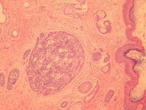
Figure 1
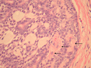
Figure 2
Special Studies for Adenoid Cystic Carcinoma
The diagnosis is fundamentally established through morphological analysis. However, certain special studies can assist in confirmation, such as PAS (Periodic Acid-Schiff) staining to highlight the basement membrane material basement membrane present in the tissue.
Differential Diagnosis of Adenoid Cystic Carcinoma
It is crucial to differentiate adenoid cystic carcinoma from other entities due to its prognosis and management. Key differential diagnoses include:
- Cylindroma: These tumors exhibit a distinctive "jigsaw puzzle" architecture, unlike the cribriform and irregular patterns of adenoid cystic carcinoma.
- Basaloid Squamous Cell Carcinoma: This can mimic the appearance of adenoid cystic carcinoma. Immunohistochemistry immunohistochemistry with p63 can be very useful in these cases. Diffuse positivity for p63 is observed more frequently in squamous cell carcinoma (squamous).scaly).
Accurate recognition of these histopathological features and the strategic use of special studies are fundamental for determining a definitive diagnosis of adenoid cystic carcinoma.


