
Actinic Granuloma: Description, Histology, and Differential Diagnosis
The Actinic Granuloma, also known as O'Brien's granuloma, is a specific dermatological condition. It is postulated that this condition arises as an inflammatory reaction inflammatory reaction secondary to extensive sun damage, particularly the progressive degeneration of the elastic fibers of the skin. Clinically, it begins by manifesting as papules pink or flesh-colored papules that, over time, undergo Fusing to form plaques of nodular to diffuse annular.
Histopathology of Actinic Granuloma
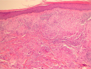
The histological examination of actinic granuloma reveals a granulomatous well-defined granulomatous reaction affecting the dermis, dermis, characterized by marked elastosis. elastosis. Detailed examinations show that the giant cells giant cells observed often contain remnants of elastic fibers, suggesting a foreign body reaction (see Figure 2). This finding is accompanied by a mixed infiltrate cellular infiltrate (Figure 1).
Pathology Findings of Actinic Granuloma
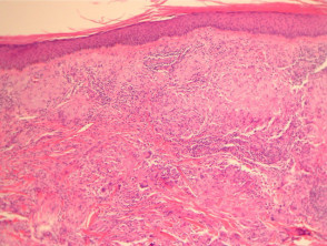
Figure 1
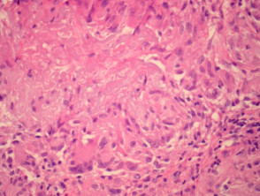
Figure 2
Special Stains for Actinic Granuloma
The application of special stains is crucial, as these techniques clearly highlight the damaged elastic fibers that have been phagocytosed by giant cells within the affected tissue.
Differential Diagnosis of Actinic Granuloma
Accurate diagnosis requires differentiating actinic granuloma from other similar conditions. It is essential to distinguish it from granuloma giant cell elastolytic. granuloma. The latter condition generally lacks the significant density of elastosis dermal dermal elastosis considered the main trigger for the granulomatous reaction in actinic granuloma. In fact, some cases have caused redundancy and confusion in nomenclature between both entities.
Another key diagnosis to rule out is Granuloma Annulare. Unlike actinic granuloma, Granuloma Annulare does not show significant elastotic material within the granulomatous reaction; instead, evidence of necrobiosis necrobiosis accompanied by a deposit of mucin in the mucin in the assessed.
Understanding the histological differences, especially in relation to dermal elastosis and the presence of foreign bodies, is essential for correctly diagnosing actinic granuloma and applying the appropriate clinical management for this manifestation of chronic sun damage.


