Calcinosis is defined as the pathological accumulation of calcium deposits in various structures, including the skin, the tissue subcutaneously, the muscle fascia, and the organs neoplasms. The most frequent are those of the gastrointestinal tract, especially when they occur in the context of the. When this manifestation primarily affects the dermal layer, it is called calcinosis cutis or, more specifically, calcificación cutaneous.
Detailed Histopathology of Calcinosis Cutis
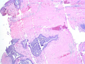
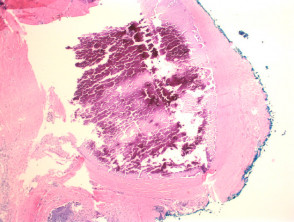
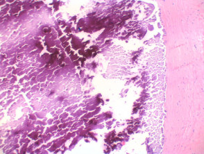
From a histological perspective, calcinosis cutis is characterized by the presence of acellular deposits that exhibit an intense affinity for basophilic stains, distributed heterogeneously within the dermis and surrounding connective tissue (see Figure 1). This basophilia is notably strong, resulting in a very pronounced purple coloration in standard stains (Figures 2 and 3).
Typically, these calcified deposits are well demarcated and often present a thin border composed of eosinophilic suggesting hyalinization. It is also common to observe an adjacent inflammatory reaction with the presence of macrophages and giant host cells or foreign body cells host cells.
A frequent artifact during histological processing is the fracturing of the calcified material and adjacent tissue, which produces artifactual lines visible in the section (Figure 1), slightly hindering morphological interpretation.
Microscopic Manifestations of Calcinosis Cutis

Specialized Histological Study Techniques
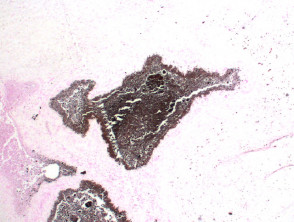
To confirm and differentiate calcium salts from other basophilic structures, special stains are used. Von Kossa silver stain is particularly useful, as it highlights mineral deposits with a characteristic black coloration (see Figure 4 for its application in calcinosis cutis).
Visualization with Von Kossa Silver Stain in Calcinosis Cutis
Understanding the histopathology and specific stains is crucial for the definitive diagnosis of calcinosis cutis, differentiating it from other conditions presenting similar deposits. The correct identification of these microscopic patterns guides subsequent clinical correlation.
Performing a Differential Diagnosis of Calcinosis Cutis
To establish an accurate diagnosis of calcinosis cutis, it is essential to differentiate it from other skin conditions that present mineral or nodular deposits. This distinction is primarily based on the histopathological analysis of the samples.
Osteoma Cutis versus Calcinosis
In the case of Cutaneous Osteoma, the key findings in the pathology are deposits showing intense eosinophilia. Furthermore, osteocytes housed within small bone lacunae are clearly distinguished, confirming the bony nature of the deposit.
Gouty Tophi: Distinctive Characteristics
toxicity Gouty Tophi are significantly differentiated by the presence of crystalline deposits. These crystals usually generate areas with a pale, often bleached appearance, and are surrounded by a marked inflammatory infiltrate composed of multinucleated giant cells. A characteristic morphological sign is the observation of needle-shaped empty spaces, arranged in a radial pattern of formation, which is indicative of urate crystals.