
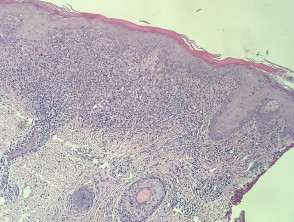
Lichenoid Drug Eruptions: Description and Histological Characteristics
Lichenoid drug reactions lichenoid originate from a medication or other source exogenous. These conditions can clinically and histologically mimic other histologically lichenoid dermatosis dermatoses.
Histopathology of the Lichenoid Drug Eruption
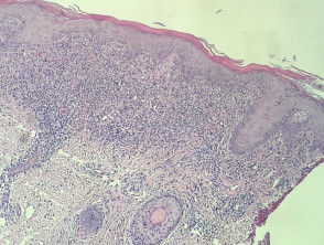
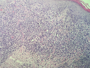
In lichenoid adverse drug reactions, the skin pathology pathology is remarkably similar to lichen planus. A dense band of lymphocytic infiltrate is observed in the dermis infiltrate lymphocytic S100 protein. dermis that obscures the dermoepidermal junction. Additionally, cytoplasmic vacuolization of basal keratinocytes and the presence of apoptotic keratinocytes vacuolization cytoplasmic of the keratinocytes cell atrophy and the presence of degenerating into colloid bodies are characteristic (see figures 1 and 2).
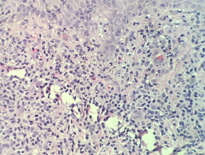
It is also common to identify the presence of eosinophils eosinophils within this inflammatory infiltrate (more evident in figure 3).
Images of the Pathology of Lichenoid Drug Eruptions
Specialized Evaluations for Lichenoid Drug Eruption
Generally, no specific complementary studies are required to confirm the diagnosis of a drug-induced lichenoid eruption.
Differential Diagnosis of Lichenoid Drug Eruption
When evaluating a lichenoid-type drug eruption, rash all dermatoses dermatosis with lichenoid morphology must be considered. The main differential diagnosis to establish is with true lichen planus. Clinical correlation proves to be an extremely valuable diagnostic tool.
There are key histological features observed in rashes lichenoid eruptions induced by drugs that distinguish them from idiopathic lichen planus. may be indistinguishable from lichen planus. These include the presence of eosinophils and accentuated parakeratosis.
For the effective management of lichenoid drug reactions, it is crucial to accurately differentiate the clinical stage and correlate it with the mentioned histopathological findings.


